BONE SCAN
A bone scan of the spine is a medical imaging test that uses a small amount of radioactive material. After insertion of the material, it uses a special camera to create pictures of the bones in the spine. The test is used to help detect and diagnose conditions such as osteoporosis, tumors, or infections in the spine. It can also be used to monitor the progress of certain treatments for these conditions.


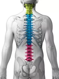
At Southwest Scoliosis and Spine Institute, we use the latest technology to diagnose spine conditions
What is a Bone Scan?
 A bone scan is a medical imaging test used to evaluate the condition of bones in the body. It involves the injection of a small amount of radioactive material, known as a radiotracer, into the bloodstream. This radiotracer accumulates in areas of the bones that are actively changing, such as those affected by injury, infection, or disease.
Why Conduct a Bone Scan?
Doctors recommend Bone Scans for various reasons, including:
- Detecting bone abnormalities: Bone scans can help identify abnormalities or changes in bone structure, especially when X-rays do not reveal small problems. They are particularly useful in detecting conditions such as fractures, stress fractures, bone infections (osteomyelitis), and bone tumors.
- Diagnosing bone metastases: Bone scans are commonly used to determine whether cancer has spread (metastasized) to the bones from another primary site. Cancer cells often accumulate in the bones, and a bone scan can reveal these areas of increased activity.
- Assessing bone healing: After a bone injury or fracture, a bone scan can monitor the healing process. It can help determine if the bone heals properly or if there are any complications, such as delayed union or nonunion.
- Evaluating bone diseases: Bone scans can aid in the diagnosis and management of various bone-related diseases, such as osteoporosis, Paget’s disease, and arthritis. They can provide information about the extent and severity of bone involvement.
- Planning orthopedic procedures: Before certain orthopedic surgeries, bone scans can evaluate the bone quality. In addition, the test can identify any abnormalities that could affect the surgical outcomes.
Questions and Answers
What is a Bone Scan?
A bone scan involves injecting a small amount of a radioactive substance, known as a radiotracer, into the bloodstream. The radiotracer accumulates in areas of the bones that are undergoing changes, such as those affected by injury, infection, or disease. A technician will then photograph the bones using a special camera that detects the radiation emitted by the radiotracer, creating detailed images of the bones.
Why is a Bone Scan necessary?
A bone scan evaluates the condition of the bones and identifies abnormalities that X-rays do not always identify. It is commonly used to detect fractures, stress fractures, bone infections, and bone tumors. Additionally, bone scans can help in diagnosing and monitoring conditions like osteoporosis, Paget’s disease, and arthritis. Doctors also use Bone Scans to assess bone healing after an injury or fracture and to determine if cancer has spread to the bones from other parts of the body.
What should I expect from a Bone Scan?
During a bone scan, a small amount of the radiotracer is injected into a vein, typically in the arm. Patients will wait for a specific period to allow the radiotracer to circulate throughout their body. Once the waiting period is over, patients will lie on a table, and a special camera will take images of their bones. The process is painless, and patients will remain still during the scan to ensure clear images. The procedure usually takes about 1-2 hours to complete. After the scan, patients can resume their normal activities, and any traces of the radiotracer will naturally leave their bodies over time. It’s important to follow any specific instructions given by your doctor before and after the scan.
Purpose of a Bone Scan
A bone scan is particularly useful for determining unexplained bone pain. By scanning the entire skeleton, doctors can diagnose a range of bone-related diseases, providing insights into issues from localized infections to systemic cancers, and everything in between.
It’s important to note that bone scans are not the first-line diagnostic tool for all bone-related conditions. Depending on the specific situation, other imaging modalities like X-rays, CT scans, or MRI scans may be preferred.
Preparing for a Bone Scan
Getting ready for a bone scan is generally straightforward, with few restrictions to keep in mind. Here are some key steps to prepare effectively:
- Inform Your Healthcare Provider: If you’ve recently taken medication containing bismuth, like Pepto-Bismol, or undergone an X-ray using barium contrast within the last four days, it’s crucial to inform your healthcare provider. These substances can affect the results of your bone scan.
- Clothing and Accessories: Opt for loose-fitting clothing on the day of your scan to ensure comfort and ease. It’s also advisable to leave jewelry and accessories at home, as you may be required to wear a gown during the procedure.
- Pregnancy and Nursing Considerations: If you’re pregnant, suspect you might be, or are currently nursing, notify your healthcare professional. Bone scans involve radiation, and precautions are taken to avoid any risk to the baby.
By following these guidelines, you’ll be well-prepared for your bone scan, ensuring the process is smooth and effective.
Considerations for Pregnant or Nursing Individuals Regarding Bone Scans
If you are pregnant or nursing, there are important considerations to keep in mind when it comes to bone scans. Typically, bone scans involve the use of radioactive tracers, which can be a concern for the developing fetus or a nursing infant due to potential radiation exposure. Here’s what you need to know:
- Pregnancy Concerns: It’s crucial to inform your healthcare provider if you are pregnant or suspect you might be. The practitioner will weigh the risks and benefits and may recommend alternative diagnostic methods that do not involve radiation, like an ultrasound or MRI.
- Nursing Considerations: If you are breastfeeding, let your healthcare provider know. Some radioactive materials used in bone scans can pass into breast milk, so your doctor might advise you to stop nursing for a certain period after the test to prevent exposure to your baby.
In general, medical professionals take great care in limiting radiation exposure, especially for those who are pregnant or breastfeeding. Always discuss your situation thoroughly with your healthcare team to ensure the safest approach tailored to your needs.
Imaging Techniques
It’s important to note that bone scans are not the first-line diagnostic tool for all bone-related conditions. Depending on the specific situation, other imaging modalities like X-rays, CT scans, or MRI scans may be preferred. The decision to conduct a bone scan is made by our doctors. We base the decision on the individual’s symptoms, medical history, and the suspected condition. In other words, a bone scan can show problems such as bone tumors, infection, and fractures of the spine. On the other hand, it can also determine bone density and the bone-thinning condition of osteoporosis.
In some cases, healthcare professionals might order additional imaging techniques to complement a bone scan. One such method is single-photon emission computerized tomography (SPECT). This advanced imaging technique can provide more detailed images, especially useful for examining bones that are deep within your body or in areas that are difficult to visualize with standard bone scans. During a SPECT scan, the camera rotates around your body, capturing comprehensive images to aid in diagnosis.
By combining these imaging methods, doctors can achieve a more complete understanding of the bone condition, ensuring a more accurate diagnosis and effective treatment plan.
The Procedure
An intravenous line (IV) is inserted into your hand or arm. Then the chemical tracer infusion takes place by going into the bloodstream through the IV. A waiting period of two to three hours will then take place, while the chemical takes effect. Basically, it attaches to any areas of bone that are undergoing rapid changes. Usually, patients can leave and come back after this period.
Patients will then lie or sit underneath a large “camera” that takes pictures of their skeleton. Since the chemical tracer is radioactive, it sends out radiation that a special camera can identify. Moreover, the camera is similar to a “Geiger counter” in that it uses film to capture the radioactivity, and the procedure takes 30-90 minutes.
Bone scans involve the use of radioactive tracers. (Mayo Clinic)
Tiny amounts of radioactive substances are injected into a vein in the hand or arm. Main images are taken 2–4 hours later to allow the tracer to circulate and be absorbed by the bones. You lie still while a camera scans you. (Mayo Clinic, 2023)
Bone Scan Risks?
There is always the risk of an allergic reaction to anything injected into the bloodstream. In this case, an allergic reaction to the chemical does not usually occur. The radioactive chemical rapidly disappears within hours from the body within hours.
Understanding the Implications of Bone Scan Results
A bone scan is a powerful diagnostic tool that provides insights into several bone-related conditions. By using nuclear imaging, this test helps detect abnormalities in bone metabolism through the use of a radioactive tracer. Here’s what the implications of bone scan results could mean for various types of cancers and bone diseases:
Cancer Detection and Monitoring
- Bone scans are pivotal in identifying cancer that has metastasized from its original site, such as the breast or prostate, to the bones.
- Results showing increased uptake may indicate areas where cancer has spread, directing further investigation and treatment.
- While less common, bone cancers originating in the bone itself can also be detected. This is crucial for early diagnosis and treatment planning.
Bone Diseases and Conditions
- Bone scans can detect both old and new fractures. This helps in assessing injuries that might not be visible on standard X-rays.
Arthritis and Degenerative Changes:
- Changes in bone metabolism might signal arthritis or other degenerative joint diseases, providing insights into the extent and activity of the condition.
Paget’s Disease of Bone:
- Increased tracer uptake can indicate abnormal bone remodeling, characteristic of Paget’s disease.
Infections:
- In cases of infections in bones or joint replacements, bone scans help pinpoint the location and severity, aiding in targeted treatments.
Understanding “Hot Spots” in a Bone Scan
In bone scans, “hot spots” appear as darker areas that signal increased activity in the bone’s metabolism. These spots are often highlighted using a radioactive tracer, making them highly visible on the scan.
What Do These Hot Spots Indicate?
Hot spots can indicate a variety of conditions, depending on their location and intensity:
- Possible Arthritis: Increased metabolic activity in joints, such as the knees, might suggest arthritis.
- Fractures: Dark areas in specific bones, like toes, could point to fractures or healing injuries.
- Cancerous Activity: Widespread hot spots may signal the presence of cancer that has spread to multiple bone sites.
- Bone Disorders: Conditions like Paget’s disease can also present as hot spots due to abnormal bone growth.
- Infections: Infected joints or bones, including those with replacements, also exhibit visible hot spots.
In conclusion, the results from a bone scan provide critical information, but they often require further evaluation to determine the exact cause of the abnormalities detected. This comprehensive analysis guides healthcare professionals in forming effective treatment strategies for a range of conditions involving the bones.
How Are the Results of a Bone Scan Interpreted?
When you undergo a bone scan, the results are meticulously analyzed by a trained professional known as a radiologist. Their expertise lies in examining the imaging results to assess unusual patterns of bone metabolism.
Hot and Cold Spots
During the scan, areas where metabolic activity deviates from the norm become visible as distinct patterns:
- Hot Spots: These areas appear darker on the scan. They indicate increased metabolic activity and are often found where tracers, the substances injected into your body, accumulate. Hot spots might suggest conditions such as fractures, infections, or tumors.
- Cold Spots: In contrast, these areas are lighter and reflect reduced tracer uptake. Cold spots can indicate issues like restricted blood flow or certain types of bone damage.
Further Investigation
While a bone scan is highly effective at detecting these metabolic anomalies, it doesn’t pinpoint their exact cause. Therefore, if hot or cold spots are identified, additional tests might be necessary. These tests could include blood tests, MRI, or CT scans to provide a comprehensive understanding of the underlying issues.
By combining these insights, your healthcare provider can gather more detailed information to guide diagnosis and treatment.
Next Steps After Discovering Abnormalities on a Bone Scan
If a bone scan reveals unusual activity or “hot spots,” it’s essential to follow up with additional steps to pinpoint the underlying issue. Here’s what you might expect next:
Further Diagnostic Tests:
- X-rays or MRI scans might be recommended to get a clearer image of the suspicious areas.
- CT scans can provide detailed cross-sectional views of bones, helping to identify fractures, infections, or tumors.
Blood Tests:
- These can detect markers of bone metabolism or infections.
- Specific tests might include measuring calcium levels or checking for signs of inflammation.
Biopsy:
- In some cases, a small tissue sample may be taken from the affected area to test for cancerous cells or other disorders.
Consultation with a Specialist:
- You might be referred to an orthopedic oncologist or a rheumatologist for expert opinions.
- This can help tailor a treatment plan specific to your needs.
Review of Medical History:
- Your doctor will likely review your medical history and any symptoms you’ve been experiencing to better understand the findings.
Further Monitoring:
- Regular follow-up scans may be scheduled to monitor any changes over time.
Keep in mind, these steps are part of a comprehensive approach to ensure accurate diagnosis and effective treatment. Discuss with your healthcare provider to determine the best course of action.
Why Choose the Southwest Scoliosis and Spine Institute
Testing is crucial in diagnosing spine problems because the spine is a complex and delicate structure that plays a vital role in supporting the body and facilitating movement. Spine-related conditions and injuries can cause a wide range of symptoms, making it challenging to pinpoint the exact cause without proper testing. Diagnostic tests such as X-rays, CT scans, and MRI scans provide detailed images of the spine, allowing healthcare professionals to assess the bones, discs, nerves, and soft tissues for any abnormalities or damage. However, in addition to the aforementioned scans, when appropriate, our doctors at the Southwest Scoliosis and Spine Institute will conduct a Bone Scan.
At the Southwest Scoliosis and Spine Institute, we focus on Diagnosis, Treatment, & Care for our Patients. Our fellowship-trained, board-certified expert orthopedic scoliosis surgeons, Richard Hostin, MD, Devesh Ramnath, MD, Ishaq Syed, MD, Shyam Kishan, MD, and Kathryn Wiesman, MD, specialize in all types of spine conditions, deformities, and scoliosis pain.
With the Bone Scan test, which is not available to all practices, our doctors, with offices in Dallas, Plano, and Frisco, Texas, can obtain accurate and comprehensive information about the spine’s condition, enabling them to make an informed diagnosis and develop an appropriate treatment plan tailored to the individual’s needs. Early and accurate diagnosis through testing is crucial for timely intervention, effective management, and improved outcomes for patients with spine problems.
____________________
Citation: Cancer.net – Bone Scans
The medical content on this page has been carefully reviewed and approved for accuracy by the Southwest Scoliosis and Spine Institute’s qualified healthcare professionals, including our board-certified physicians and Physician Assistants. Our team ensures that all information reflects the latest evidence-based practices and meets rigorous standards of medical accuracy, with oversight from our expert spine doctors to guarantee reliability for our patients.
If you or a loved one suffers from spinal pain, you owe it to yourself to call Southwest Scoliosis and Spine Institute at 214-556-0555 to make an appointment.