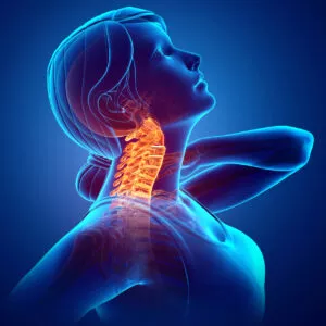
CERVICAL HERNIATED DISC
At Southwest Scoliosis and Spine Institute, our board-certified, fellowship-trained orthopedic specialists have extensive experience treating complex spine conditions, including herniated cervical discs. A herniated cervical disc can lead to severe neck pain, radiating arm pain, and even numbness or weakness, impacting daily activities and overall quality of life.
Our Cervical Herniated Disc Doctors are dedicated to providing accurate diagnosis and customized treatment options, including minimally invasive approaches, to help patients find relief, restore function, and regain comfort in their daily lives.


Herniated discs also occur in the neck (cervical spine) and, more rarely, in the upper back (thoracic spine).
Cervical Herniated Disc
 At Southwest Scoliosis and Spine Institute, our board-certified, fellowship-trained orthopedic doctors Richard Hostin, MD, Devesh Ramnath, MD, Ishaq Syed, MD, Shyam Kishan, MD, and Kathryn Wiesman, MD have treated thousands of patients with complex spine conditions that included cervical herniated discs. A herniated disc, also known as a slipped or ruptured disc, refers to damage to the intervertebral discs that cushion the vertebrae in the spine. The intervertebral discs comprise a tough outer layer called the annulus fibrosus and a soft inner core called the nucleus pulposus.
When the annulus fibrosus becomes weakened or torn, the nucleus pulposus can bulge out or rupture, causing the disc to press against nearby nerves or the spinal cord.1 This can lead to pain, numbness, or weakness in the arms, legs, or back. While a herniated cervical disc can occur from injury or trauma, the effects of aging cause many more cases.
Because this ailment occurs over time, patients often don’t notice the symptoms until they become severe. Also, this ailment typically occurs between the ages of 30 and 50. Unfortunately, it tends to affect older adults because age causes tissue to become less effective. Therefore, cervical discs crack, and other injuries occur to increase the risk of herniation.
Questions and Answers
What are the Symptoms of a Cervical Herniated Disc
The symptoms of a cervical herniated disc can vary depending on the location and severity of the herniation. Common symptoms include pain in the neck, shoulders, and arms, numbness or tingling in the arms or hands, and weakness in the arms or hands. Some people may also experience headaches, dizziness, or difficulty with balance.
How do doctors diagnose a Cervical Herniated Disc
Doctors typically diagnose a cervical herniated disc through a combination of physical examination, imaging tests such as MRI or CT scans, and nerve function tests. Your doctor may also ask about your medical history and any other existing symptoms.
What are the Non-Surgical Treatments for a Cervical Herniated Disc
Non-surgical treatments for a cervical herniated disc may include physical therapy, pain management, and the use of cervical traction or cervical collars to support the neck. In some cases, steroid injections will reduce inflammation and relieve pain.
Cervical Herniated Disc
“Cervical Herniated Disc means that the gel between your vertebra in your neck is coming out. Symptoms are usually neck and arm pain. Treatments are medications, physical therapy exercise, targeted injections and lastly, surgery.”
Cervical Herniated Disc Information
Numerous cases of cervical herniated discs can indeed go undiagnosed. This often happens because symptoms can appear mild or mistaken for other common ailments. Diagnostic rates may also differ depending on the population being studied, leading to variations in estimates.
The estimated rate of cervical herniated discs in adults typically falls from 0.5% to 2%.2
When it comes to the risk of developing a cervical herniated disc, the jury has been out for quite some time. Historically, men were believed to suffer the condition more than women. However, recent research indicates that women might face a higher risk. This evolving understanding suggests that both genders must practice good cervical spine health, although the risk factors may differ.
Anatomical Components of a Cervical Disc
Understanding the anatomy of a cervical disc involves looking at its two primary components: the annulus fibrosus and the nucleus pulposus.
Annulus Fibrosus
The annulus fibrosus serves as the disc’s robust outer layer. Composed of concentric rings made of collagen fibers, this structure provides the necessary strength to protect the softer interior. Its key role includes managing the spine’s heavy loads and absorbing shocks from everyday activities. In summary, the annulus fibrosus gives the disc its durability and shock-absorbing capabilities, while the nucleus pulposus adds cushioning and flexibility. These two components work together to ensure the cervical disc can handle both stress and movement efficiently.
Nucleus Pulposus
Nestled within the annulus fibrosus is the nucleus pulposus, a gel-like core. This inner component consists of a network of fibers suspended in a mucoprotein gel, which allows it to act as a cushion. This cushioning effect provides both flexibility and support for spinal movements.
Understanding the Impact of Different Disc Levels on Motor Deficits, Reflex Changes, and Sensory Deficits
The alignment of your spinal discs plays a crucial role in maintaining overall nervous system function. When there’s a problem at specific disc levels, it can affect various areas of your body differently. Here’s a breakdown of how different disc levels can influence motor deficits, reflex changes, and sensory deficits:
C4-5 Disc Level
- Motor Deficits: You might experience weakness in your deltoid muscles, which are responsible for lifting and rotating your shoulder.
- Reflex Changes: Generally, there are no significant reflex changes noted at this level.
- Sensory Deficits: Sensation loss or numbness may appear on the lateral side of your shoulder.
C5-6 Disc Level
- Motor Deficits: Weakness in your biceps can occur, affecting your ability to bend your elbow and rotate your forearm.
- Reflex Changes: Changes in the bicep reflex are commonly observed.
- Sensory Deficits: You could notice sensory loss or numbness extending down the lateral part of your arm and forearm, including the thumb and index finger.
C6-7 Disc Level
- Motor Deficits: Weakness in your triceps and wrist extensors can occur, causing challenges in extending your arm and wrist.
- Reflex Changes: Triceps reflex alterations are typically seen.
- Sensory Deficits: Sensation changes or numbness might affect your middle finger.
C7-T1 Disc Level
- Motor Deficits: You may face difficulties with hand grip and wrist flexion, impacting your ability to grasp objects and bend your wrist.
- Reflex Changes: Reflex changes are usually not noted at this level.
- Sensory Deficits: A sensory deficit might occur along the ring and small fingers, affecting tactile sensation.
Key Takeaways
- Motor Deficits: Different disc levels impact various muscle groups, such as deltoids (C4-5), biceps (C5-6), and triceps (C6-7).
- Reflex Changes: Not all disc levels result in reflex changes, but when they do, common areas include the biceps and triceps.
- Sensory Deficits: Sensory impairment is often localized to specific regions, like the lateral shoulder at C4-5 and fingers at C7-T1.
Understanding these correlations can help in diagnosing and managing the symptoms associated with spinal disc issues. If you experience any of these symptoms, consider consulting a healthcare professional for a thorough evaluation.
The Causes of a Herniated Disc:
- Age: As we get older, the discs in our spine lose water content, making them less flexible and more prone to injury.
- Repetitive stress: Repeated lifting, twisting, or bending can put stress on the discs and increase the risk of herniation.
- Poor posture: Sitting or standing in a slouched position can place undue pressure on the discs and increase the risk of injury.
- Obesity: Excess weight can put added stress on the spine and increase the risk of herniation.
- Genetics: Some people may get herniated discs because of genetic factors.
- Trauma: A sudden impact, such as a fall or car accident, can cause a herniated disc.
- Bulging disc in the neck
Symptoms of a Cervical Herniated Disc
To clarify, herniated disc symptoms can differ based on the location of the disc and the compression of any nerves. In this case, symptoms often include:
- Pain in the neck that radiates down the arm towards the hands and fingers
- Numbness or tingling in the shoulders, arms, or hands
- Muscle weakness in the hand and/or arms
In severe cases, a herniated disc may put pressure on the spinal cord. This will cause pain, tingling, numbness, or weakness in both arms and possibly even lower in the body.
Symptoms of spinal cord compression may also include:3
- Difficulty walking straight or an unsteady gait
- A reduction in fine motor skills in the hands and arms
- Painful tingling or feelings like electric shocks in the torso or even into the legs.
Understanding Myelopathy: Causes and Symptoms
Myelopathy refers to a condition where the spinal cord becomes compressed, often due to issues such as cervical disc herniation. This compression can lead to a range of serious symptoms.
Symptoms of Myelopathy
- Difficulty Walking: Patients often experience challenges with mobility and feel unstable or uncoordinated.
- Spasticity: An increase in muscle stiffness and involuntary spasms.
- Bowel and Bladder Incontinence: Loss of control over bladder and bowel functions is a significant indicator.
These symptoms result from the pressure placed on the spinal cord, disrupting normal neurological functions. If you notice any of these signs, it’s crucial to seek medical attention promptly.4
Herniated Disc Diagnosis
Your doctor will review your medical history. Then he or she will perform a thorough physical exam and look for any signs of limited mobility. After that, the doctor will ask about any pain, check balance, muscle reflexes, loss of sensation, or muscle weakness. If doctors suspect a cervical herniated disc, they will use an X-ray to confirm the diagnosis.
Because X-rays can only show issues with the bones, the doctor may order a CT or MRI scan. For imaging, we use a state-of-the-art digital low-dose X-ray system. In less than a minute, this advanced unit takes high-quality patient images. Also, this system can take X-rays of patients in a standing or seated position. Moreover, it’s located inside our Dallas office, and you and your doctor can review the X-rays ASAP.
How do Doctors Assess Sensory Loss in Patients with a Suspected Herniated Disc?
Assessing sensory loss in patients with a suspected herniated disc involves a multifaceted approach. Here’s how medical professionals typically conduct these evaluations:
Muscle Strength Testing
Each muscle group is evaluated individually to identify any signs of weakness. This step is crucial, as weakened muscles can indicate nerve damage or compression.
Sensory Examination
Doctors use fine touch and light pinpricks to examine the entire body. These tests help detect subtle loss of sensation. By comparing the results from different areas, practitioners can pinpoint regions affected by nerve issues.
Reflex Testing
Reflexes are tested at key points such as the elbows, hands, knees, and ankles. Abnormal reflexes in these areas can provide clues about the location of a herniated disc. For example, a diminished knee reflex might indicate an issue in the lumbar spine. Muscle strength tests are crucial in diagnosing a herniated disc. By evaluating the strength of specific muscle groups, healthcare providers can identify areas of weakness that may indicate nerve impingement. This is essential in pinpointing which nerves are affected by the disc herniation.
In addition to muscle strength tests, physicians often use fine touch and light pin examination across the body. This helps to detect subtle sensory loss, which can indicate nerve damage. Reflex tests at various joints, such as elbows, hands, knees, and ankles, are also conducted. Abnormal reflexes in these areas can reveal the precise location of the herniated disc. Together, these tests provide a comprehensive view of how a herniated disc is impacting the nervous system, offering targeted insight for effective treatment strategies. Using these methods, healthcare providers can accurately assess sensory loss and identify the underlying causes, ensuring appropriate treatment plans are devised.
Understanding Radiculopathy and Its Connection to Cervical Herniated Discs
Radiculopathy refers to a condition characterized by pain, numbness, or weakness that radiates along the path of a specific nerve. When it occurs in the neck region, it often stems from issues with the cervical discs. These discs serve as cushions between the vertebrae in the spine. When one of these discs herniates, it can press on spinal nerves, leading to radiculopathy.
Symptoms of Radiculopathy
- Neck Pain: Begins in the neck and may extend down to the shoulder.
- Arm and Hand Pain: Radiates down the arm and into the hand, following the path of the affected nerve.
- Numbness and Weakness Can affect the arm and hand, making everyday tasks challenging.
Link to Cervical Herniated Discs
Cervical herniated discs occur when the gel-like center of a disc bulges out through a tear in the tougher outer layer. This herniation can compress nearby nerves, causing the symptoms associated with radiculopathy.
Other Possible Complications
- Spinal Cord Compression: In severe cases, a herniated disc may also compress the spinal cord, leading to myelopathy. This condition can cause significant issues with walking, muscle stiffness (spasticity), and even control over bladder and bowel functions.
Understanding this relationship is crucial for accurate diagnosis and treatment. If symptoms of radiculopathy are present, addressing the underlying cervical disc issue often provides relief and prevents further complications.
Treatment for a Cervical Herniated Disc
As a result, treatment options range from physical therapy and medicines to surgery. At our offices, our doctors always start with a conservative approach to treatment before thinking about surgery. In addition, our Doctors always take the time to discuss all options with our patients. So we talk with the patient about our options and how treatment depends on age, health, and severity of the problem.
Non-surgical Treatment
Most patients see rapid improvement with medicines that reduce pain, inflammation, and muscle stiffness that accompany a herniated disc. Medicines manage pain, while numbness or tingling tends to improve with time and rest. The initial treatment for a herniated cervical disc is usually conservative. This often involves non-steroidal anti-inflammatory medication such as ibuprofen, Aleve, or Motrin, along with rest. By combining these methods, many patients experience significant relief and a return to normal activities without the need for more invasive procedures. Non-surgical treatments for a Cervical herniated disc include:
- Physical therapy and/or exercises to relieve the pressure on the nerves
- Medications to reduce swelling and pain, such as non-steroidal anti-inflammatory drugs (NSAIDs) like ibuprofen, Aleve, and Motrin
- Epidural steroid injections and nerve root injections reduce swelling around the affected disc and nerves, as well as relieve acute pain radiating to the hips or down the legs.
Doctors can conservatively manage approximately 80% of herniated discs with the above methods.5 Physical therapy often employs techniques such as traction, ultrasound, and electrical muscle stimulation to relax muscles that are in spasm and secondarily inflamed from the compressed spinal nerve. In some cases, doctors will suggest an epidural steroid injection using a spinal needle under X-ray guidance to ensure the medication is directed precisely to the level of the disc herniation.
Epidural steroid injection
An epidural steroid injection utilizes a spinal needle under X-ray guidance to direct the medication to the exact level of the disc herniation. This precise targeting helps ensure the medication effectively reduces inflammation and pain. A specialist typically performs the procedure to ensure it is done safely and accurately.6
Cervical Herniated Disc Surgery
If the pain, numbness, and other symptoms persist for more than 6-12 weeks, or if evidence suggests a severe spinal cord compression, your doctor may recommend surgery.
Surgical Treatment Includes:
Anterior Cervical Discectomy and Spinal Fusion involves removing the bad disc through the front of the neck. This will relieve the pressure on the nerves and/or spinal cord. Because most (if not all) of the disc gets removed, a fusion procedure will then stabilize the spine. A fusion procedure involves a small bone graft, screws, and rods to ensure that the bones fuse properly. This procedure, known as Anterior Cervical Discectomy and Fusion (ACDF), begins with an incision in the neck to expose the front of the spine.
The damaged disc is then carefully removed to alleviate pressure on the spinal cord and nerve roots. Following the removal, a bone graft is placed in the space left by the disc. The graft can come either from a bone bank or the patient’s hip. To further support the spine, a metal plate bridges the area, ensuring stability during the healing process. It’s also worth noting that multiple-level ACDFs can occur if more than one disc is affected. This adaptability makes ACDF a versatile solution for various degrees of cervical spine issues.
Posterior access
Posterior Cervical Decompression (Microdiscectomy). In cases where the herniation of the disc appears minor, your doctor may use a minimally invasive technique. The Doctor removes the portion of the disc pressing on the nerve and makes a small incision on the back of the neck. Because only the small herniated portion of the disc gets removed, this surgery does not usually need to include a spinal fusion procedure. Posterior Cervical Discectomy. During this operation, bone is removed from the back of the spine to expose the compressed nerve root.
The disc is then removed, creating space for the nerve root to pass through. This procedure is designed to alleviate pressure on the nerve, providing relief from pain and other symptoms. By combining these techniques, surgeons can tailor the approach to the patient’s specific needs, ensuring the best possible outcome with the least invasive methods.
Understanding Artificial Discs in Cervical Disc Disease Treatment
Artificial discs are advanced medical implants designed to replace damaged or degenerated cervical discs in the spine. These prosthetic devices mimic the natural structure and function of a healthy spinal disc, providing an innovative solution for patients suffering from cervical disc disease.
Disc arthroplasty
Disc arthroplasty, also known as disc replacement surgery, is a procedure designed to treat conditions like herniated discs and degenerative disc disease by replacing a damaged intervertebral disc in the spine with an artificial implant. This surgery typically involves making a small incision to access the affected disc, removing the problematic tissue, and inserting a prosthetic device made of materials such as metal and polyethylene. The artificial disc is engineered to preserve natural spinal movement, allowing for flexibility in the back or neck while alleviating pain and pressure on nerves. Performed under general anesthesia, the operation usually takes 1-3 hours, depending on the location and complexity, with patients often experiencing a relatively quicker recovery compared to spinal fusion due to the maintenance of mobility.
One of the primary advantages of disc arthroplasty is its ability to reduce the risk of adjacent segment degeneration, as it maintains the spine’s natural biomechanics rather than fusing vertebrae together. It is most commonly applied in the cervical or lumbar regions for patients with chronic pain who have not responded to conservative treatments like physical therapy or medications. However, potential risks include infection, implant failure, or nerve damage, which are evaluated on a case-by-case basis. Overall, the success of the procedure depends on factors such as the patient’s age, activity level, and the specific type of implant used, making it a promising option for eligible individuals seeking long-term relief and improved quality of life.
How Are Artificial Discs Used?
When treating cervical disc disease, a surgeon removes the problematic disc and inserts the artificial disc into the disc space. Here’s a breakdown of the key benefits and aspects of this procedure:
- No Bone Graft Required: Unlike traditional spinal fusion, the installation of an artificial disc doesn’t necessitate a bone graft. This simplifies the surgical procedure and reduces the recovery time.
- Maintains Mobility: A significant advantage of artificial discs is their ability to preserve the natural motion of the neck. This contrasts with fusion surgery, which can limit flexibility and movement.
- Prevents Adjacent Disc Disease: By retaining normal neck function, artificial discs may help prevent the premature degeneration of adjacent discs. This is a common issue in patients who undergo traditional spinal fusion, where the added stress on nearby discs accelerates wear and tear.
Why Choose Artificial Discs?
Patients with cervical disc disease often experience chronic pain and mobility issues that can severely impact their quality of life. Artificial disc replacement types provides a promising alternative to fusion surgery, offering benefits such as:
- Quicker Recovery: Reduced need for post-operative immobilization and potentially shorter rehabilitation periods.
- Enhanced Quality of Life: Maintaining natural spine movement can significantly improve daily activities and overall well-being.
- Potential for Reduced Future Interventions: By minimizing the risk of adjacent disc disease, patients may face fewer additional surgeries or treatments down the line.
In conclusion, artificial discs represent a significant advancement in the treatment of cervical disc disease, providing patients with an option that supports both recovery and long-term spinal health.
Understanding Corpectomy and Fusion vs. ACDF
When it comes to treating herniated cervical discs, two common surgical options are available: Anterior Cervical Discectomy and Fusion (ACDF) and Corpectomy and Fusion. Both procedures aim to alleviate pain and restore spinal stability, but they differ in terms of their approach and scope.
Anterior Cervical Discectomy and Fusion (ACDF)
Procedure:
- ACDF involves a small incision at the front of the neck to access the spine.
- The surgeon will remove the damaged disc carefully, which helps relieve pressure on the spinal cord and adjacent nerve roots.
Bone Graft and Stabilization:
- After disc removal, the surgeon will insert a bone graft with a metal plate to ensure stability.
- The bone graft comes from either a donor bank or the patient’s hip.
Multiple Levels:
- Surgeons can use this technique to treat multiple levels of the spine if several discs are problematic.
Corpectomy and Fusion
- In contrast to ACDF, a corpectomy involves not just removing the damaged disc but also part or all of the affected vertebral body.
Procedure:
- The surgeon removes a large section of the bone, which provides more extensive decompression of the spinal cord and nerves.
Bone Graft and Metal Plate:
- Similar to ACDF, surgeons use a bone graft and metal plate to bridge the gap and stabilize the spine.
Key Differences
Extent of Removal:
- ACDF focuses on removing only the herniated disc, while corpectomy goes a step further by removing a section of the vertebral bone.
Indications:
- Surgeons typically perform an ACDF when one or a few discs are involved, whereas corpectomy occurs for more severe cases requiring broader decompression.
Surgical Impact:
- Corpectomy may involve a longer recovery due to the more extensive nature of the surgery.
Understanding these differences helps in making informed decisions about which surgical option aligns with the patient’s condition. Please always consult a specialized spine surgeon to determine which procedure best fits your medical needs.
Why Choose the Southwest Scoliosis and Spine Institute with Offices in Dallas, Plano, and Frisco, TX
There are many reasons why you should choose the Southwest Scoliosis and Spine Institute doctors to treat your lumbar spinal scoliosis. Here are a few:
- A team of experienced and board-certified surgeons who specialize in treating spinal conditions, including scoliosis.
- They offer a variety of treatment options, including non-surgical and surgical treatments.
- A state-of-the-art facility that maintains the latest technology for diagnosing and treating spinal conditions.
- A reputation throughout the Nation for providing the very best spinal care for their patients.
- They are conveniently located in three locations in Dallas, Plano, and Frisco, Texas.
If you are considering treatment for lumbar spinal scoliosis, we encourage you to schedule a consultation with the Southwest Scoliosis and Spine Institute. They can help you to understand your condition and develop a personalized treatment plan for your condition.
____________________
Citations:
- National Library of Medicine. Cervical Disc Herniation. StatPearls.
- National Institute of Neurological Disorders and Stroke. Cervical Myelopathy.
- American Academy of Orthopaedic Surgeons. Cervical Radiculopathy (Pinched Nerve).
- National Library of Medicine. Epidemiology of Cervical Disc Herniation.
- American Academy of Orthopaedic Surgeons. Non-Surgical Treatment of Herniated Disc.
- National Library of Medicine. Cervical Epidural Steroid Injection. StatPearls.
The medical content on this page has been carefully reviewed and approved for accuracy by the Southwest Scoliosis and Spine Institute’s qualified healthcare professionals, including our board-certified physicians and Physician Assistants. Our team ensures that all information reflects the latest evidence-based practices and meets rigorous standards of medical accuracy, with oversight from our expert spine doctors to guarantee reliability for our patients.