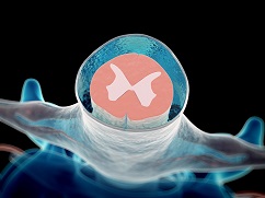
SPINAL TAP
Spinal tap refers to both a procedure and a lab test. Furthermore, Spinal fluid is extracted from the lower back and then tested to determine the spinal fluid pressure and contents.


 Could you tell me why the Spinal Tap Procedure is being conducted?
Doctors will perform a spinal tap to get a sample of cerebrospinal fluid (the fluid that surrounds the spinal cord) to test the pressure and content of the fluid. The fluid is typically very clear and contains proteins, sugars, and other substances found in blood. And, it typically does not contain red blood cells or many white blood cells. In addition, signs of problems include evidence of bleeding, an increase in white blood cells (infection-fighting cells), and an increase in protein levels or inflammation. Moreover, this could mean there is an infection, tumor, or hemorrhage around the brain or spinal cord.
How Safe is a Spinal Tap Procedure Overall?
Spinal taps, also known as lumbar punctures, are generally considered safe procedures with complications being quite rare. The most common issue patients might encounter is a headache, which occurs in about 10% to 30% of cases. This headache typically develops several hours or even a day or two after the procedure. While these headaches can be quite uncomfortable, they are not dangerous and do not pose any neurological risks.
Tips to Manage Headache:
- Hydration: Drinking plenty of fluids such as water, coffee, tea, or soda can help prevent and alleviate headaches.
- Pain Relievers: Over-the-counter pain medications can offer relief.
- Consult Your Doctor: If the headache persists beyond two days, it’s advisable to contact your healthcare provider to rule out any severe issues.
Overall Safety
The key takeaway is that spinal taps are very safe procedures when performed by skilled medical professionals. The vast majority of individuals undergoing this procedure experience little to no discomfort or anxiety. With proper technique and care, complications are infrequent, ensuring a generally positive outcome for most patients.
Don’t let fear deter you—a spinal tap is a safe and well-regulated procedure designed to provide important diagnostic information while minimizing risk.\
Is a Spinal Tap a Dangerous Procedure?
Spinal taps, or lumbar punctures, are generally considered safe medical procedures. The most common side effect is a headache, experienced by about 10% to 30% of patients. These headaches typically manifest several hours or even days after the procedure and, while uncomfortable, are not harmful.
Rare but Possible Severe Complications
Though rare, more severe complications can occur. These include:
- Infection
- Bleeding
- Numbness
- Brain herniation (movement of brain tissue due to pressure)
Importantly, nerve or spinal cord damage is extremely rare.
Expert Opinion
Experts emphasize that fear of the procedure is often unfounded. When performed by skilled professionals, spinal taps are both safe and gentle. Complications are uncommon, and the procedure should cause minimal discomfort and anxiety.
In summary, while no medical procedure is without risk, the likelihood of severe complications from a spinal tap is very low.
When Might Someone Need a Spinal Tap?
A spinal tap, or lumbar puncture, is a crucial medical procedure that serves multiple diagnostic and therapeutic purposes. Here are some common scenarios where a doctor will order a spinal tap:
Diagnosing Infections
Spinal taps are frequently performed to identify infections in the central nervous system (CNS). By extracting a sample of cerebrospinal fluid (CSF), medical professionals can detect the presence of infectious organisms, aiding in the diagnosis of conditions such as meningitis. The test results help tailor antibiotic treatments effectively.
Identifying CNS Disorders
The procedure can diagnose several CNS disorders, including:
- Multiple Sclerosis
- Guillain-Barré Syndrome
- Epilepsy
Cancer Diagnosis
Spinal taps can also detect cancers affecting the brain or spinal cord, providing valuable information that guides treatment plans.
Therapeutic Uses
- Chemotherapy Administration: For certain cancers, doctors can administer chemotherapy drugs directly into the CSF.
- Anesthesia: Doctors can deliver anesthetics via a spinal tap for specific surgical procedures.
Imaging Needs
In some cases, contrast agents (dyes) are injected into the CSF to enhance imaging of the spinal cord and its coverings, especially useful when MRI scans are not possible.
Procedure Settings and Urgency
A spinal tap can occur in a hospital or an outpatient facility, depending on the urgency and purpose. While it is not considered an emergency procedure, it is viewed as urgent and typically performed within hours rather than minutes.
A spinal tap is a versatile medical procedure offering crucial insights and treatments for an array of conditions, solidifying its role in modern medicine.
Preparing for a Spinal Tap
Preparing for a lumbar puncture test involves a few important steps to ensure the procedure goes smoothly:
- Follow Pre-Test Instructions: Adhere to any guidelines given by your healthcare provider regarding food, drink, or medications. You might need to stop consuming certain substances, particularly blood thinners, before the test.
- Discussion with Medical Staff: The doctor or nurse will inform you about the procedure. They will explain potential risks, address any questions you might have, and ask you to sign consent forms.
- Preparatory Medications: Your doctor may administer a mild sedative or give you intravenous fluids to help you stay calm during the procedure. Your doctor may provide other medications as necessary.
In addition to these steps, make sure to inform your healthcare team about any drug allergies you have. Depending on the specific instructions from your provider, you may need to refrain from eating or drinking for a few hours before the procedure. Wear comfortable clothing, as you will change into a gown upon arriving at the hospital.
On the morning of your appointment, contact your doctor if you experience any unusual symptoms or feel unwell. Ensure you have arranged for someone to drive you home after the procedure, as you might feel a bit weak or dizzy afterward.
What Happens During a Spinal Tap
A lumbar puncture, often known as a spinal tap, is a medical procedure used to access the cerebrospinal fluid (CSF) surrounding the brain and spinal cord. Here’s how it works:
After changing into the gown and becoming settled, your back is washed with antiseptic soap or iodine and covered with a sterile drape to create a clean field. Then, the patient lies on their side with knees drawn up to the chest, or sits and leans forward.
“We want your knees pulled up as far as possible and your chin down to your chest so you’re curled up into a little ball,” explains Dr. Hostin. This position helps arch your back and spaces out your vertebrae, giving the doctor a larger target area.
For infants or young children, someone, usually a parent or a nurse, may help hold them in the right position during the procedure. In some cases, parents are allowed to stay in the room for support. It’s always a good idea to discuss this in advance with your doctor.
A local anesthetic is injected into your lower back to numb the puncture site before the needle is inserted. There’s a brief sting as the anesthetic goes in, but it quickly dulls the area.
“Then, the needle itself gets inserted into the skin between the bones in the lower back, and into the spinal space where the cerebrospinal fluid lives,” says Dr. Hostin. In certain situations, an ultrasound or special X-ray imaging technique called fluoroscopy is used to determine the best position for the spinal needle, especially for young children or those with challenging anatomy.
What the patient feels
You might feel some pressure in your back as the needle advances, but most people describe it as more odd than painful. Occasionally, the doctor may ask you to adjust your position slightly to help with needle placement.
Further Tests
In addition to diagnostics, a lumbar puncture can administer treatments. For example, it can deliver contrast dyes needed for imaging tests like a myelogram, inject anesthetics to numb the spinal cord, or dispense medications to alleviate conditions such as hydrocephalus. Each of these steps, from positioning the patient to collecting CSF or administering treatments, is conducted with precision to ensure the best possible outcome and patient safety.
Dr. Hostin notes, “We’ll collect a teaspoonful, usually—it’s all we really need. And then the needle comes out. Put a little Band-Aid on the skin, and we’re all done.” It’s crucial to remain still during the procedure to help complete it faster and more efficiently. Although spinal tap pain is rare, sometimes the needle may brush by a nerve root, causing a brief zing or electric shock sensation down one leg.
Once the lumbar puncture test is complete, the doctor will gently press on the puncture site to prevent any bleeding. A bandage will cover the area to keep it clean and protected.
The entire process generally takes about 45 minutes from start to finish. Afterward, your healthcare provider may suggest you lie down for a short while to minimize the risk of a headache and help your body recover. Sometimes, especially for babies and young children, an ultrasound will guide the needle safely and prevent it from going in too far.
Throughout, your comfort and safety are top priorities—whether you’re the patient or the parent standing by.
How is a Spinal Tap Performed?
A lumbar puncture, often known as a spinal tap, is a medical procedure used to access the cerebrospinal fluid (CSF) surrounding the brain and spinal cord. Here’s how it works:
First, the patient lies on their side with knees drawn up to the chest, or sits and leans forward to increase the space between the vertebrae. A healthcare professional then sterilizes the lower back area to minimize infection risk.
Next, a hollow needle is carefully inserted through the skin of the lower back, between the bones of the spine, and into the spinal canal. This is the space where the cerebrospinal fluid flows around the spinal cord.
Once the needle is in place, the doctor will collect CSF for various diagnostic tests. This fluid helps detect conditions such as infections, bleeding around the brain, or multiple sclerosis. The procedure can also measure CSF pressure to check for hydrocephalus, a condition characterized by an accumulation of CSF in the brain.
In addition to diagnostics, a lumbar puncture can administer treatments. For example, it can deliver contrast dyes needed for imaging tests like a myelogram, inject anesthetics to numb the spinal cord, or dispense medications to alleviate conditions such as hydrocephalus. Each of these steps, from positioning the patient to collecting CSF or administering treatments, is conducted with precision to ensure the best possible outcome and patient safety.
Post-Procedure Care
Once the lumbar puncture test is complete, the doctor will gently press on the puncture site to prevent any bleeding. A bandage will cover the area to keep it clean and protected.
- It’s important to take it easy. Avoid any heavy lifting or strenuous activities for the next 24 hours to aid in your recovery.
- You will lie on your back for 30 to 60 minutes immediately following the procedure so your doctor can monitor for any adverse effects.
Hydration:
- Drink plenty of fluids. Staying well-hydrated helps replenish the cerebrospinal fluid that was collected.
Monitoring the Puncture Site:
- Keep an eye on the bandaged area. If you notice any blood or fluid leaking, notify your doctor immediately.
Pain Management:
- Pain medication can usually address any discomfort you may feel post-procedure.
Activity Resumption:
- If your lumbar puncture was an outpatient procedure, you may leave the facility and resume simple activities after taking a few hours to relax. However, this can depend on the reason for the procedure. For example, if there is a concern for conditions like meningitis, you may need to stay in the hospital for further observation.
Follow-Up and Results:
- Speak with your provider about when you can expect results. It can take anywhere from a day up to a week, depending on why you received the lumbar puncture.
To make the most of your follow-up appointment, consider writing down questions you want to ask your healthcare professional. Don’t hesitate to bring up any concerns or new questions that arise during your visit. Some helpful questions might include:
- Based on the results, what are my next steps?
- What kind of follow-up, if any, should I expect?
- Are there any factors that might have affected the results of this test and, therefore, may have altered the results?
- Will I need to repeat the test at some point?
Following these steps will help ensure a smooth recovery after your lumbar puncture test. For questions, we invite our patients to call us at the Southwest Scoliosis and Spine Institute.
Managing Post-Spinal Tap Headaches
Here are some effective strategies to manage these headaches:
- Hydration: Drinking plenty of fluids such as water, coffee, tea, or soda may help prevent or alleviate the headache.
- Over-the-Counter Pain Relievers: Medications like acetaminophen or ibuprofen can provide relief.
- Rest: Taking it easy and lying down for a while may also help minimize discomfort.
If your headache persists for more than two days, it’s important to contact your healthcare provider. This could indicate a more serious issue that may require medical attention.
What to Do If a Headache Persists After a Spinal Tap
Experiencing a headache after a spinal tap is relatively common and often resolves on its own. However, if the headache lasts more than two days, it’s crucial to take action.
Steps to Take:
- Monitor Your Symptoms: Keep a close eye on how you’re feeling. Note down any additional symptoms such as nausea, dizziness, or visual disturbances.
- Stay Hydrated: Drink plenty of fluids, especially those high in caffeine, like coffee or tea, as they can help relieve spinal headaches.
- Rest: Lie down as much as possible, as this can alleviate headache severity.
- Contact Your Healthcare Provider: After two days, if the headache continues or worsens, reach out to your doctor. Prolonged headaches could indicate a more serious issue needing immediate medical attention.
- Discuss Treatment Options: Your doctor might suggest various treatments, such as a blood patch, where a small amount of your blood is injected into the spinal canal to seal any leaks causing the headache.
By following these steps, you can manage prolonged headaches effectively and ensure any underlying issues are promptly addressed.
What is the Prognosis for Someone with a Herniated Thoracic Disc?
The outlook for individuals with a herniated thoracic disc is generally favorable, especially when the condition is identified early and managed appropriately. Most people respond well to conservative treatments such as rest, medication, and physical therapy, experiencing significant relief from pain and other symptoms. In many cases, these nonsurgical approaches are all that’s needed.
Occasionally, if symptoms are severe or do not improve with standard therapies, surgical intervention might become necessary. When surgery is recommended—often due to ongoing pain or progressing neurological changes—many patients notice significant improvement, including relief from pain and, in some cases, recovery of lost function.
It’s important to note that many thoracic disc herniations are discovered incidentally during imaging for unrelated concerns, and they may never result in pain or noticeable symptoms. When this occurs, doctors typically recommend observation rather than immediate treatment, stepping in only if new symptoms arise.
Severe complications like paralysis are rare and tend to develop slowly, giving patients time to seek medical care before any permanent disability occurs. Most individuals receive treatment long before the condition advances to serious nerve damage. While surgery can often reverse gradually developing symptoms, there’s still a possibility that some issues may persist.
As always, it’s crucial to consult a healthcare provider if you notice any new or worsening symptoms, as early intervention can make a meaningful difference in long-term recovery.
Anatomy of the Mid-Back
To understand your symptoms and treatment choices, patients should understand the anatomy of the mid-back. As such, patients should become familiar with the various parts that constitute the thoracic spine and how they work together.
The spine is made up of 33 vertebrae, divided into five distinct segments running from the base of your skull down to your pelvis:
- Cervical: the neck region
- Thoracic: the middle back (mid-back)
- Lumbar: lower back
- Sacrum: connects the spine to the hips
- Coccyx: the tailbone
The thoracic spine comprises 12 vertebrae, extending from the base of your neck to the bottom of your rib cage. What sets the thoracic vertebrae apart from those in other regions is their direct connection to the ribs. Each thoracic vertebra has joints that attach to a rib on either side, providing additional support and stability. This rib cage support means the thoracic spine experiences less wear and tear compared to the cervical and lumbar segments, making conditions like thoracic herniated discs less common in this region.
Learn more about the anatomy of the thoracic spine.
The intervertebral discs refer to the shock-absorbing cushions between each vertebra of your spine. For instance, only one disc exists between each vertebra. Meanwhile, each disc has a strong outer ring of fibers, called the annulus, and a soft, jelly-like center, called the nucleus pulposus. The annulus refers to the disc’s outer layer and the strongest area of the disc. Also, the annulus helps connect each vertebra. The nucleus in the center of the disc serves as the main shock absorber.
A herniated disc occurs when the intervertebral disc’s outer fibers (the annulus) become damaged, and the soft inner material of the nucleus pulposus ruptures out of its normal space. If the annulus tears near the spinal canal, the nucleus pulposus material can push into the spinal canal.
Causes of a Thoracic Herniated Disc
Herniated discs can occur in children, although rarely. However, a true herniated nucleus pulposus usually happens in young and middle-aged adults and generally occurs in the lower back. Additionally, disc herniations in the thoracic spine mostly affect people between the ages of 40 and 60. In older folks, the degenerative disc changes that occur in the spine with aging make it less likely for them to suffer a true herniated disc. Thoracic herniated discs typically arise from degenerative changes in the spine, which are often linked to the aging process. However, several other factors can also contribute to the development of this condition.
- Trauma and Injuries: A sudden injury or acute trauma to the spine, such as from an accident, can cause a disc in the thoracic region to herniate. Improper lifting techniques or handling heavy objects can also strain the spine, leading to herniation.
- Repetitive Strain: Occupations or activities involving repetitive movements can put continuous stress on the spine, increasing the likelihood of disc herniation. Over time, this constant strain weakens the spinal discs.
- Genetic Factors: Herniated discs can sometimes run in families. Genetic predisposition may play a role, making some individuals more susceptible to this condition.
- Lifestyle Choices: Certain lifestyle factors can elevate the risk of disc herniation. Smoking, for instance, has been linked to accelerated disc degeneration, while excess body weight puts additional pressure on the spinal discs, making herniation more likely.
Additional causes
- Other Contributing Factors: Age-related spinal degeneration is a primary cause, but it’s crucial to note that having any of these risk factors does not guarantee the development of a thoracic herniated disc.
- Disc Degeneration: One of the primary culprits is disc degeneration. As we age, the spinal discs lose their moisture content and elasticity. This natural wear and tear makes the discs more prone to herniating, particularly in the thoracic region.
- Obesity: Carrying excess weight puts additional pressure on the spine. This added strain can accelerate disc degeneration and heighten the risk of herniation.
- Poor Posture: Lastly, poor posture, especially from sitting for extended periods, can misalign the spine and place uneven stress on the thoracic discs. By being aware of these causes, you can take proactive steps to mitigate the risk and seek treatment if needed. If you’re experiencing persistent upper back pain, consult a healthcare professional to explore the best course of action.
Understanding these causes can help in taking preventive measures to protect your spine from unnecessary strain and injury.
How Common Is A Herniated Disc In The Thoracic Region Of The Spine?
A herniated disc in the thoracic (upper) region of the spine is quite uncommon. Most disc herniations typically occur in the lower back or the cervical (neck) area. When a herniation does take place in the thoracic region, it often leads to upper back pain, which can sometimes extend to the chest or abdomen.
The thoracic spine is uniquely protected compared to other parts of the spine. Each thoracic vertebra is joined to a rib on either side, creating a sturdy rib cage that provides additional support and stability. This extra support means the thoracic region experiences less wear and tear, making it less likely to develop herniated discs or other degenerative conditions. In fact, thoracic herniated discs account for less than 1 percent of all herniated discs, making them quite rare. The thoracic spine also has the highest number of vertebrae, but is the least mobile region, further reducing its susceptibility to disc herniation.
In rare cases, a thoracic herniated disc may press against the spinal cord, leading to serious conditions like myelopathy, a type of spinal cord dysfunction. This situation is a medical emergency and requires prompt attention. Overall, while herniated discs appear in other parts of the spine, they rarely occur in the thoracic region.
Discs
Discs can also rupture from a small amount of force, usually due to the weakening of the annulus from repeated injuries that add up over time. As the annulus becomes weaker, at some point lifting or bending causes too much pressure across the disc. Then, the weakened disc ruptures while doing something that five years earlier would not have caused a problem. Furthermore, the effects of aging comprise the most common reason for disc herniation in the thoracic spine.
The ruptured material can enter the spinal canal from the nucleus pulposus and can cause pressure on the nerves in the spinal canal. Also, there is some evidence that the nucleus pulposus material causes a chemical irritation of the nerve roots. Both the pressure on the nerve root and the chemical irritation can lead to problems with how the nerve root functions. Thus, the combination of the two can cause pain, weakness, and numbness in the area of the body to which the nerve supplies sensation.
Understanding Radiculopathy and Its Connection to Thoracic Herniated Discs
What is Radiculopathy?
Radiculopathy refers to a condition where a spinal nerve becomes compressed, leading to a variety of symptoms such as pain, numbness, and tingling sensations that extend from the spine along the affected nerve pathway. This condition can significantly impact your daily life, depending on the severity and location of the nerve compression.
How Does a Thoracic Herniated Disc Cause Radiculopathy?
In the context of a thoracic herniated disc, radiculopathy occurs when the herniation exerts pressure on a spinal nerve within the thoracic region of the spine. This type of herniation happens when the soft inner gel of a spinal disc protrudes through its tougher exterior. Here’s how it affects you:
- Pain and Discomfort: You may experience sharp or radiating pain that wraps around your rib cage or upper abdomen, following the path of the affected nerve.
- Numbness and Tingling: These sensations often accompany the pain, adding to the discomfort and potentially affecting your ability to carry out routine tasks.
- Localized Symptoms: While thoracic herniated discs are less common than cervical or lumbar herniations, the symptoms can be particularly disruptive due to the central location in the spine.
Recognizing these symptoms early and seeking appropriate medical advice can help manage the condition and reduce its impact on your quality of life.
Thoracic Herniated Disc Symptoms
Whether your symptoms stem from a herniated thoracic disc or another spinal condition, it’s important to seek professional evaluation. Your primary care doctor can help rule out other possible causes of your pain and, if appropriate, refer you to a specialist for further assessment. A consultation with a spine surgeon or spinal neurosurgeon can clarify whether surgery or a conservative treatment approach would best address your unique situation.
Taking that first step toward expert care ensures you receive a thorough diagnosis and a treatment plan tailored to your needs—so you can get back to living life with less pain and more confidence.
Identifying symptoms of a thoracic herniated disc is crucial for timely diagnosis and treatment. These symptoms vary based on the location and severity of the herniation. Here’s what you need to look out for:
Central Disc Protrusion
- Upper Back Pain: This is a typical symptom when the disc herniates centrally. The pain may intensify based on the size of the herniation and the pressure it exerts on the spinal cord.
- Myelopathy: A more severe symptom, characterized by spinal cord dysfunction, which can manifest as difficulty walking, loss of coordination, or even incontinence.
- Paralysis: In critical cases, significant pressure on the spinal cord can lead to paralysis below the waist.
Lateral Disc Herniation
- Radiating Pain: When the disc herniates to the side, it can press on the nerve root exiting the spine, causing pain that may radiate to the chest wall or abdomen.
- Localized Discomfort: Pain may occur around the area of the herniation, depending on which nerves are affected.
Centro-Lateral Disc Herniation
- Combination of Symptoms: This type of herniation can present a mix of the above symptoms, such as upper back pain, radiating pain, or myelopathy. The specific symptoms depend on the extent and direction of the protrusion.
When it comes to herniated discs, the thoracic vertebrae most commonly impacted are T11 and T12. In fact, the majority of these cases—around 75%—occur below the T8 vertebra. This proximity to the more flexible lumbar spine might contribute to their susceptibility. Understanding these symptoms can aid in recognizing a potential thoracic herniated disc early on, thereby enabling more effective management and care.
____________________
Citation: Wikipedia -Spinal Tap
The medical content on this page has been carefully reviewed and approved for accuracy by the Southwest Scoliosis and Spine Institute’s qualified healthcare professionals, including our board-certified physicians and Physician Assistants. Our team ensures that all information reflects the latest evidence-based practices and meets rigorous standards of medical accuracy, with oversight from our expert spine doctors to guarantee reliability for our patients.
What are the Limitations?
The spinal tap does not provide information about most types of back pain. It is not helpful if your doctor suspects you may have arthritis of the spine, a herniated disc, or spinal stenosis.
What are the Risks?
A spinal tap—also known as a lumbar puncture—has more risks associated with it than most other tests. This is one reason that doctors prefer to use “noninvasive” tests first, such as MRI & CT-scan. The risks associated with a spinal tap include meningitis (infection of the spinal fluid) and the possibility of developing a spinal headache. However, there is a very small chance that the needle will cause bleeding around the spinal sac.
This is more of a risk if you are on medications that thin the blood and interfere with blood clotting. Therefore, if possible, you should not take aspirin or ibuprofen for five days before having this test. In addition, make sure to tell your doctor if you are taking any type of blood thinners.
While these are relatively common issues, there are some more severe complications to be aware of. Although extremely rare, a spinal tap can lead to:
- Infection: Beyond meningitis, there is the possibility of more generalized infections.
- Bleeding: Severe bleeding around the spinal sac or even within the spinal cord itself.
- Numbness: This can occur if the nerves are affected by the procedure.
- Brain Herniation: The movement of brain tissue due to pressure changes, which is a critical condition.
Nerve or spinal cord damage is also extremely uncommon but worth noting. Understanding these risks is crucial for making an informed decision about undergoing a spinal tap. Always consult with your healthcare provider to weigh the benefits and risks based on your specific medical condition.
Why Is a Spinal Tap Done?
Despite these risks, a spinal tap is sometimes necessary because it provides important information that other tests can’t. Your doctor may order a lumbar puncture to:
- Collect cerebrospinal fluid to check for infections, inflammation, or other diseases.
- Measure the pressure of the cerebrospinal fluid.
- Inject spinal anesthetics, chemotherapy, or other medications directly into the fluid around your spinal cord.
- Inject contrast dye or radioactive substances to help create diagnostic images of the fluid’s flow (procedures known as myelography and cisternography).
The results from a spinal tap can help diagnose a range of serious conditions, including:
- Infections: Bacterial, viral, or fungal infections such as meningitis, encephalitis, or syphilis.
- Bleeding: Bleeding around the brain (subarachnoid hemorrhage).
- Certain Cancers: Cancers that involve the brain or spinal cord.
- Nervous System Disorders: Autoimmune and inflammatory conditions like multiple sclerosis or Guillain-Barré syndrome.
- Neurological Conditions: Some forms of dementia, including Alzheimer’s disease.
So, while there are risks involved, the information gained from a spinal tap can be vital in diagnosing and treating potentially life-threatening conditions. Always discuss your options and concerns with your healthcare provider to make the most informed choice.
What Should I Do If I Experience a Spinal Fluid Leak After a Lumbar Puncture?
If you suspect a spinal fluid leak after a lumbar puncture, follow these essential steps to ensure your health and safety:
- Rest and Limit Activity: Make sure to rest and avoid any strenuous activities for at least 24 hours following the procedure. This helps your body recover and reduces the risk of complications.
- Stay Hydrated: Drink plenty of fluids to stay hydrated. Hydration can aid in your recovery and help maintain proper fluid balance in your body.
- Monitor the Puncture Site: Keep a close eye on the puncture site. If you notice any blood or fluid leaking, it’s critical to inform your doctor immediately.
By following these guidelines and maintaining clear communication with your healthcare provider, you can effectively manage your recovery and address any issues promptly.
If you have additional questions about the lumbar puncture test, it’s important to reach out directly to the physician who ordered it by calling our office at the Southwest Scoliosis and Spine Institute.
How is Cerebrospinal Fluid (CSF) Pressure Measured During a Lumbar Puncture?
To measure cerebrospinal fluid (CSF) pressure during a lumbar puncture, medical professionals follow a detailed process. Initially, the patient is asked to straighten their legs. This position helps to reduce abdominal pressure, thereby allowing for a more accurate assessment of the CSF pressure.
A specialized needle is then carefully inserted into the spinal canal. Once in place, the needle is connected to a pressure-measuring device known as a manometer. This device records the CSF pressure, providing essential data about the conditions within the spinal canal.
Essentially, the combination of the patient’s positioning and the use of a precise measuring instrument ensures an accurate determination of CSF pressure during this procedure.
The typical range for cerebrospinal fluid (CSF) pressure lies between 70 and 180 millimeters of mercury (mmHg).
To receive your lumbar puncture test results, the initial step involves the doctor examining the color of your cerebrospinal fluid (CSF). Typically, CSF is clear, but color variations can provide immediate insights. For instance:
- A reddish hue might indicate bleeding or a subarachnoid hemorrhage.
- A cloudy or yellowish appearance could point to an infection, such as meningitis.
Beyond color, your CSF sample is thoroughly analyzed in a laboratory for a range of important details:
- General appearance: In addition to color, clarity is noted. Cloudiness may signal elevated white blood cells or protein, both signs of infection or inflammation.
- Protein levels: Normally, total protein in CSF is below 45 milligrams per deciliter (mg/dL). Higher levels may suggest infections, inflammatory conditions, or other neurological issues.
- White blood cell count: CSF typically contains up to five white blood cells per microliter. A higher count can indicate infection or inflammation.
- Glucose (sugar) levels: Low glucose in the CSF may point to an infection, tumor, or certain other conditions.
- Presence of microorganisms: Bacteria, viruses, or fungi found in the CSF are strong indicators of infection.
- Abnormal cells: Detection of tumor cells or immature blood cells in the CSF can assist in diagnosing certain cancers or blood disorders.
All these findings are combined with information gathered during the procedure, such as CSF pressure, to help your doctor make an accurate diagnosis. Typically, you’ll receive your results within a few days, but some specialized tests may take a little longer. If you’re anxious about the timing, don’t hesitate to ask your healthcare provider when to expect your results.
What Information Does a Lumbar Puncture Provide?
A lumbar puncture, also known as a spinal tap, reveals a wealth of critical information about your health by analyzing cerebrospinal fluid (CSF). This fluid, which surrounds the brain and spinal cord, can indicate a range of conditions.
Common Findings from a Lumbar Puncture
- Infections: By examining the CSF, doctors can diagnose infections such as meningitis, which affects the membranes covering the brain and spinal cord, or encephalitis, a viral infection of the brain.
- Bleeding: Detection of blood in the CSF helps identify serious issues like a subarachnoid hemorrhage or stroke.
- Tumors: The presence of abnormal cells can signal tumors, including cancers like lymphoma.
- Autoimmune Disorders: Diseases such as multiple sclerosis can be detected through specific markers found in the CSF.
Additional Insights
Beyond looking for abnormal cells, a lumbar puncture also measures CSF pressure. Normal CSF pressure ranges between 70 and 180 mm H2O. Deviations from this range can indicate conditions like hydrocephalus, where there is excessive accumulation of cerebrospinal fluid.
In summary, a lumbar puncture provides comprehensive data that aids in diagnosing infections, bleeding, tumors, autoimmune disorders, and abnormal pressure conditions.
____________________
Citation: National Institute of Health – Spinal Tab
The medical content on this page has been carefully reviewed and approved for accuracy by the Southwest Scoliosis and Spine Institute’s qualified healthcare professionals, including our board-certified physicians and Physician Assistants. Our team ensures that all information reflects the latest evidence-based practices and meets rigorous standards of medical accuracy, with oversight from our expert spine doctors to guarantee reliability for our patients.
If you or a loved one suffers from spinal pain, you owe it to yourself to call Southwest Scoliosis and Spine Institute at 214-556-0555 to make an appointment.