ANATOMY OF THE SPINE
The spine consists of up to 24 bones, called vertebrae. Ligaments and muscles connect these bones together to form the spinal column. Also, the spinal column gives the body form and function and protects the spinal cord. (Hostin et al., 2016a)
At the Southwest Scoliosis and Spine Institute, our expert Spine Doctors and Surgeons are dedicated to diagnosing and treating spinal problems in children and adults to ensure comprehensive care tailored to each patient’s needs. With advanced techniques and a compassionate approach, our team diagnoses, treats, and cares for patients suffering from Spinal Conditions.


Our Doctors, Surgeons, and Staff want you to know about the Spine. We hope that the information below is helpful.
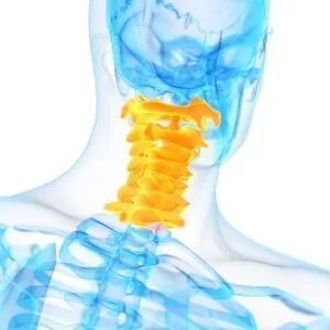
Anatomy of the Spine – Cervical Spine (Neck )
The cervical vertebrae, making up the section of the spine corresponding to the neck, are uniquely designed, comprising seven distinct bones. Notably, the first two vertebrae deviate from the typical spinal structure. The first, known as the atlas (C1), is ring-shaped and supports the skull, allowing the head to pivot. The second, called the axis (C2), features a peg-like projection, around which the atlas rotates, facilitating head movement. (Standring, 2020)
Apart from these two, the remaining five cervical vertebrae (C3 to C7) share a common structure with most spinal segments, including intervertebral discs at the front and facet joints at the back. However, a distinguishing characteristic of these vertebrae is the presence of transverse foramina—holes in each vertebra that allow for the passage of arteries to the brain, notably the vertebral arteries.
Additionally, the cervical vertebrae play a crucial role in safeguarding the spinal cord. The spinal cord runs through a canal in the center of these vertebrae, connecting the brain to various parts of the body. This not only facilitates neural communication but also provides vital protection to the spinal cord itself.
The anatomy of the spine includes the cervical spine, which consists of the first seven vertebrae in the spine. It starts just below the skull and ends just above the thoracic spine. The cervical spine possesses a lordotic curve, a backward “C” shaped just like the lumbar spine. In addition, the cervical spine’s mobility helps both of the other spinal regions. Think about all the directions and angles you can turn your neck.
 Arteries in the Cervical Spine
Unlike the rest of the spine, special openings in each vertebra in the cervical spine contain arteries (blood vessels that carry blood away from the heart). The cervical spine, known for its exceptional mobility compared to other spinal regions, features unique anatomical characteristics. Notably, each vertebra in this area has special openings designed specifically for arteries. (Moore et al., 2018)
These are not just any arteries; they are pivotal conduits that carry blood away from the heart. Additionally, the arteries that run through these openings bring blood to the brain, fulfilling a critical role in maintaining brain health and function.
This intricate arrangement allows for the efficient supply of oxygen and nutrients essential for cerebral operations, highlighting the sophisticated nature of human anatomy in supporting our body’s complex systems.
Vertebrae
Two vertebrae in the cervical spine, the atlas and the axis, differ from the other vertebrae because they facilitate rotation. These two vertebrae are the reason your neck can move in so many directions.
The atlas refers to the first cervical vertebra, and its location appears between the skull and the rest of the spine. Additionally, the atlas does not have a vertebral body, but it does have a thick forward (anterior) arch and a thin back (posterior) arch with two prominent sideways masses.
In addition, the atlas sits on top of the second cervical vertebra, the axis. The axis contains a bony knob called the odontoid process, which sticks up through the hole in the atlas. Special ligaments between the atlas and the axis allow for a great deal of rotation. Also, this unique situation allows the head to turn from side to side as far as it can.
The cervical spine is very flexible, but it is also very much at risk for injury from strong, sudden movements, such as whiplash-type injuries. This high risk of harm occurs due to the limited muscle support that exists in the cervical area, and the fact that this part of the spine supports the weight of the head, which weighs an average of 15 pounds.
This appears as a lot of weight for a small, thin set of bones and soft tissues to bear. Accordingly, sudden, strong head movements can cause damage.
Anatomy of the Spine – Thoracic Spine (Mid Back)
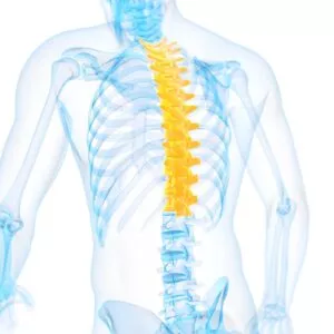 The thoracic spine appears unique among other segments of the spine because pairs of rib bones extend from the spaces between its vertebrae. The ribs’ curved shapes create a cage-like structure that houses and protects many vital organs, including the heart and lungs. Also, the thoracic spine, which comprises the upper and middle back, includes twelve vertebrae identified as T1 to T12.
Unlike the more flexible cervical spine in the neck or the lumbar spine in the lower back, the thoracic spine exhibits limited movement.
This restricted motion primarily occurs due to its integral connection with the ribs and sternum. Because these bones are firmly linked, they provide a sturdy framework that limits the range of mobility but also significantly decreases the likelihood of injuries or degenerative issues in this mid-back area. Essentially, the ribcage acts as a protective barrier and anchor, stabilizing the spine in this region.
Because the body moves so much in daily life, the thoracic spine can get injured for many reasons, from improper posture to a compression fracture. Some injuries can put pressure on the spinal nerves, creating even stronger pain and other symptoms.
The thoracic spine exhibits a kyphotic curve, a “C”-shaped curve with the opening facing forward. This part of the spine has very narrow, thin intervertebral discs. Rib connections and smaller discs in the thoracic spine limit the amount of spinal movement in the mid-back compared to the lumbar or cervical parts of the spine.
Anatomy of the Spine – Lumbar Spine (Low Back )
Another part of the anatomy of the spine, the lowest part of the spine, is called the lumbar spine. Additionally, this area usually has five vertebrae. However, sometimes people are born with a sixth vertebra in the lumbar region. Also, the base of your spine (called the sacrum) consists of a group of specialized vertebrae that connect the spine to the pelvis.
Sacrum – The main function of the sacrum connect the spine to the hip bones (iliac). There are five sacral vertebrae, which are fused. Together with the iliac bones, they form a ring called the pelvic girdle.
When one of the bones forms as a vertebra rather than part of the sacrum, doctors refer to it as a transitional (or sixth) vertebra. Nevertheless, this occurrence does not appear to have any serious side effects.
The lumbar spine’s shape has a lordotic curve-shaped like a backward “C”. If you think of the spine as having an “S” shape, the lumbar region’s location can be found at the bottom of the “S”. Also, the vertebrae in the lumbar spine area are the largest in the entire spine.
Conversely, the lumbar spinal canal looks larger than in the cervical or thoracic parts of the spine, and as such, the size of the lumbar spine allows for more space for nerves to move about.
Low back pain occurs in lots of people because the lumbar spine connects to the pelvis, where most of your weight-bearing and body movement takes place. Typically, people tend to place too much pressure on this area, such as when lifting a heavy box, twisting to move a heavy load, or carrying a heavy object. Consequently, these activities can cause repetitive injuries that can lead to damage to the parts of the lumbar spine.
The Cauda Equina
The cauda equina refers to a bundle of nerves located at the lower end of the spinal cord, known as the conus medullaris. This collection of nerves plays a crucial role in transmitting signals between the lower body and the brain, affecting motor control and sensation in the legs and pelvic organs.
The name “cauda equina” comes from the Latin term for “horse’s tail.” Early anatomists named it this because the nerve bundle’s appearance resembles the tail of a horse, spreading out in a similar, hair-like manner.
Key Points:
- Location: At the base of the spinal cord
- Function: Transmits motor and sensory signals to the lower body
- Appearance: Resembles a horse’s tail, hence the name “cauda equina”
Understanding the cauda equina remains essential for grasping how our nervous system manages critical functions in our lower extremities.
Anatomy of the Spine
Your spine consists of 24 small bones, called vertebrae. The vertebrae protect and support the spinal cord. In addition, they also bear the majority of the weight put upon your spine. Normally, vertebrae, like all bones, have a hard and strong outer shell, called cortical bone. The inside consists of a soft, spongy type of bone, called cancellous bone.
The vertebral body consists of a large, round portion of the bone. Each vertebra attaches to a bony ring. When the vertebrae are stacked one on top of the other, the rings create a hollow tube for the spinal cord to pass through. Each vertebra attaches to the others by groups of ligaments. Ligaments connect bones to bones, like tendons connect muscles to bones. Equally, some tendons fasten muscles to the vertebrae.
To further understand spine anatomy, it’s essential to familiarize yourself with some key terms:
Glossary of Spine Anatomy Terms
- Annulus fibrosus: The fibrous, ring-like outer portion of an intervertebral disc.
- Anterior: Refers to the front of the body or given structure.
- Anterolateral: Situated or occurring in front of and to the side.
- Arachnoiditis: Inflammation of the arachnoid membrane (the middle of the three protective layers called the meninges); most commonly seen around the spinal cord and cauda equina.
- Arthritis: Inflammation of a joint, usually accompanied by swelling, pain, and restriction of motion.
- Bone spur: Bony growth or rough edge of bone.
- Cauda equina: The collection of nerves at the end of the spinal cord that resembles a horse’s tail.
- Cervical spine: The neck region of the spine, consisting of the first seven vertebrae.
- Coccyx: More commonly known as the tailbone, this appears as a bony structure in the region of the spine below the sacrum.
- Conus medullaris: The cone-shaped bottom of the spinal cord, usually at the level of L1.
- Disc (Intervertebral): A tough, elastic cushion located between the vertebrae in the spinal column; acts as a shock absorber for the vertebrae.
Glossary Continued
- Disc degeneration: The deterioration of a disc. A disc in the spine may wear out over time. A deteriorated disc may or may not cause pain.
- Distal: Located downstream.
- Facet: A joint formed when a posterior structure of a vertebra joins with a facet of an adjacent vertebra; this joint allows for motion in the spinal column. Each vertebra has a right and left superior (upper) facet and a right and left inferior (lower) facet.
- Foramen: An opening in the vertebrae of the spine through which the spinal nerve roots travel.
- Herniated disc: A Condition in which the jelly-like core material of a disc bulges or ruptures out of its normal position; a herniated disc may exert pressure on the surrounding nerve root and/or the spinal cord.
- Joint: The junction of two or more bones that permits varying degrees of motion between the bones.
- Lamina: The flattened or arched part of the vertebral arch that forms the roof or back part of the spinal canal.
- Lateral: Situated on the side or away from the midline of the body.
- Ligament: Fibrous connective tissue that links bones together at joints or that passes between bones of the spine.
- Lumbar spine: The lower back region of the spine; it consists of the five vertebrae between the ribs and the pelvis.
The Glossary Continued
- Nerves: Neural tissue that conducts electrical impulses (messages) from the brain and spinal cord to all other parts of the body; also conveys sensory information from the body to the central nervous system.
- Nerve root: The initial portion of a spinal nerve as it originates from the spinal cord.
- Neural arch: The bony arch of the back part of a vertebra that surrounds the spinal cord; also referred to as the vertebral arch, it consists of the spinous process and lamina.
- Pedicle: The bony part of each side of the neural arch of a vertebra that connects the lamina (back part) with the vertebral body (front part).
- Posterior: The back or rear side of the body or a given structure.
- Proximal: Located upstream.
- Rotation: Twisting movement of one vertebra on another as a patient turns from one side to the other.
- Sacrum: Part of the pelvis just above the coccyx (tailbone) and below the lumbar spine (lower back).
- Sciatica: A lay term indicating pain along the course of the sciatic nerve; typically noted in the back of the buttocks and running down the back of the leg and thigh to below the knee.
- Scoliosis: An abnormal sideways curvature of the spine.
Glossary Continued
- Spinal canal: A bony channel located in the vertebral column that protects the spinal cord and nerve roots.
- Spinal cord: The longitudinal cord of nerve tissue enclosed in the spinal canal. It serves not only as a pathway for nerve impulses to and from the brain but also as a center for operating and coordinating reflex actions independent of the brain.
- Spinal stenosis: Abnormal narrowing of the vertebral column that may result in pressure on the spinal cord, spinal sac, or nerve roots stemming from the spinal cord.
- Spine: The flexible bone column extending from the base of the skull to the tailbone. It consists of 33 bones known as vertebrae, and doctors refer to this as the vertebral column, spinal column, or backbone.
- Spondylitis: Inflammation of the vertebrae.
- Spondylolisthesis: The forward displacement or “slippage” of one vertebra onto another.
- Spondylosis: Degenerative bony changes in the spine, usually most marked at the vertebral joints and intervertebral discs.
- Superior: Situated above or directed upward toward the head of an individual.
- Thoracic spine: The region of the spine attached to the ribcage; located between the cervical and lumbar areas, it consists of 12 vertebrae.
- Vertebrae: The 33 bones that make up the spine, individually referred to as a vertebra. They are divided into the cervical spine (neck), the thoracic spine (upper back or rib cage), the lumbar spine (lower back), and the sacral spine (pelvis or base of the spine).
Understanding these terms will provide a solid foundation for comprehending the complexities of spine anatomy and related conditions.
How Do Intervertebral Discs Change With Age?
As we age, our intervertebral discs undergo several changes that impact their functionality and overall health.
Loss of Fluid Reabsorption
One significant change is the discs’ reduced ability to reabsorb fluid. This reduction leads to the discs becoming less pliable and more brittle. Essentially, they lose the sponginess that helps cushion our vertebrae, becoming flatter and less effective at absorbing shocks experienced during daily activities.
Development of Bone Spurs
Additionally, age-related diseases such as osteoarthritis and osteoporosis can contribute to the formation of bone spurs, also known as osteophytes. These bony projections can develop on the edges of the vertebrae, further complicating spinal health and potentially causing discomfort or pain.
Risk of Bulging and Herniation
With age, the risk of disc bulging or herniation increases. Due to gradual wear and tear, combined with potential injury or strain, the inner gel-like nucleus of the disc may push through the outer annulus. This herniation can compress nearby nerve roots, leading to back pain and other symptoms.
Key Points:
- Reduced Fluid Reabsorption: Discs become less flexible and more brittle.
- Formation of Bone Spurs: Osteoarthritis and osteoporosis contribute to additional bone growth on the vertebrae.
- Increased Risk of Bulging/Herniation: Wear and tear, along with injuries, can cause the disc’s nucleus to protrude, compressing nerves.
Understanding Disc Degeneration
Disc degeneration refers to the wear and tear of the discs in your spine. These discs act as cushions between the vertebrae, allowing for flexibility and absorbing shock. Over time, the annulus fibrosus (the tough outer layer) may weaken, allowing the nucleus pulposus (the soft inner core) to either bulge or completely escape through the annulus fibrosus. This can cause nerve or spinal cord compression, leading to pain.
The Process of Degeneration
- Early Life: In the initial years, spinal discs have a blood supply that keeps them nourished and healthy.
- Adulthood (20s and 30s): The blood supply gradually diminishes, and the discs become avascular, meaning they no longer receive direct blood flow.
- Middle Age (50 and beyond): By the age of 50, over 95% of individuals experience some degree of disc degeneration. The discs lose water content and shrink, which reduces their ability to support the spine and absorb shocks.
Effects of Disc Degeneration
As the discs degenerate, several issues may arise:
- Reduced Flexibility: The spine loses some of its range of motion.
- Decreased Shock Absorption: The spine becomes less effective at absorbing impacts.
- Pain: The degenerated discs can generate pain either through nerve compression or by affecting the surrounding vertebrae.
Prevalence by Age 50
Disc degeneration is incredibly common as people age. By the time individuals reach the age of 50, it affects more than 95% of them, making it a nearly ubiquitous condition among older adults. In summary, disc degeneration is a natural aging process of the spinal discs, impacting nearly everyone in their 50s and leading to potential spinal issues and pain. Understanding these changes can help in managing age-related spinal issues and maintaining overall spinal health.
Vertebra structure
Understanding the structure of a vertebra is essential for grasping how our spine functions. Each vertebra, despite its unique regional variations, shares three primary functional components:
- Body: This drum-shaped section is built to bear the body’s weight and resist compression. It acts as the main support pillar within the vertebra.
- Vertebral Arch: This arch-shaped element shelters the spinal cord, providing crucial protection. It forms a protective tunnel through which the spinal cord passes.
- Processes: These star-shaped projections serve as attachment points for muscles. The processes extend outward, acting as levers that muscles can pull on to support movement and stability.
By breaking down the vertebra into these three functional parts—the body, the vertebral arch, and the processes—we gain a clearer understanding of its critical role in our skeletal system.
Intervertebral Disc
Between each vertebra, a soft, gel-like cushion called an intervertebral disc exists. These flat, round “cushions” act like shock absorbers by helping absorb pressure. The discs prevent the bones from rubbing against each other. Altogether, each disc has a strong outer ring of fibers called the annulus, and a soft, jelly-like center called the nucleus pulposus.
The annulus is the strongest area of the disc. It helps keep the disc’s center intact. The annulus is a strong ligament that connects each vertebra.
A mushy nucleus of the disc serves as the main shock absorber. Furthermore, the nucleus consists of moist tissue because it has high water content. The water content helps the disc act like a shock absorber-somewhat like a waterbed mattress.
Facet Joints
A person’s spinal column has real joints (just like the knee, elbow, etc.) called facet joints. The facet joints link the vertebrae together and give them the flexibility to move against each other. The facets are the “bony knobs” that meet between each vertebra. There are two facet joints between each pair of vertebrae, one on each side. They extend and overlap each other to form a joint between the neighboring vertebral facet joints. Lastly, the facet joints give the spine flexibility.
Facet joints are synovial joints, structures that allow movement between two bones. In this case, the ends of the bones that make up a synovial joint are covered with articular cartilage, a slick, spongy material that allows the bones to glide against one another without much friction.
Moreover, the synovial fluid inside the joint keeps the joint surfaces lubricated, like oil lubricates the parts of a machine. Furthermore, this fluid is contained inside the joint by the joint capsule, a watertight sac of soft tissue and ligaments that fully surrounds and encloses the joint.
Neural Foraminal
The spinal cord branches off into 31 pairs of nerve roots, which exit the spine through small openings on each side of the vertebra called neural foramina. (Netter, 2019)
The two nerve roots in each pair go in opposite directions when traveling through the foramina. One goes out through the left foramina, and the other goes out through the right foramina. Then the nerve root allows nerve signals to travel to and from your brain to the rest of your body.
Spinal Cord
The spinal cord contains a column of millions of nerve fibers that carry messages from your brain to the rest of your body. It is located immediately below the brain stem and extends through the foramen magnum, a hole at the base of the skull, down to the area between the end of your first lumbar vertebra and the top of your second lumbar vertebra.
Each vertebra has a hole in the center, so when they stack on top of each other, they form a hollow tube (spinal canal) that holds and protects the entire spinal cord and its nerve roots.
The spinal cord functions as a sophisticated network, carrying information from the outer elements of the body (skin, muscles, ligaments, joints) through the sensory tracts to the central “computer,” the brain. Data are processed there, and new information, such as muscle control, is sent out through the motor tracts of the spinal cord.
The spinal cord is divided into thirty-one pairs of spinal nerves. The spinal nerves function as “telephone lines,” relaying messages between your body and the spinal cord to control sensation and movement. Each spinal nerve consists of two roots. Additionally, the ventral (front) root transports motor impulses from the brain, while the dorsal (back) root carries sensory impulses to the brain.
The ventral and dorsal roots fuse to form a spinal nerve that travels down the spinal canal alongside the cord until it reaches its exit hole, the intervertebral foramen. To understand more precisely, let’s break down the spinal nerves by their regions and exit points:
Cervical Nerves
- 8 pairs of cervical nerves exit the cervical cord at each vertebral level. The first cervical root exits above the C1 vertebra. The second cervical root exits between the C1-C2 segment, and the remaining roots exit just below the corresponding numbered vertebra. The eighth nerve root exits between the C7 and T1 vertebrae.
Thoracic Nerves
- 12 pairs of thoracic nerves: The first nerve root exits between the T1 and T2 vertebrae.
Lumbar Nerves
- 5 pairs of lumbar nerves: The first of these nerve roots exits between L1 and L2.
Sacral Nerves
- 5 pairs of sacral nerves: The first nerve root exits between S1 and S2.
Coccygeal Nerves
- 1 pair of coccygeal (Co1) nerves meets in the area of the tailbone.
This detailed breakdown helps to fully understand the organization and exit points of the spinal nerves, illustrating how the nervous system intricately connects various parts of the body to the spinal cord and brain. The spinal cord only goes down to the second lumbar vertebra. Below this level, the spinal canal contains a group of nerve fibers, called the cauda equina syndrome.
This group of nerves goes to the pelvis and lower limbs. A protective membrane, called the dura mater, covers the spinal cord. The dura mater forms a watertight sac around the spinal cord and the spinal nerves. Inside this sac, the spinal cord is surrounded by spinal fluid.
Nerve Roots
First, the nerve fibers in your spinal cord branch off to form pairs of nerve roots that travel through the small openings between your vertebrae. Notably, the nerves in each area of the spinal cord connect to specific parts of your body. As a result, damage to the spinal cord can cause paralysis in certain areas and not others. It ultimately depends on which spinal nerves are affected.
For instance, the nerves of the cervical spine go to the upper chest and arms. Similarly, the nerves of the thoracic spine go to the chest and abdomen, and the nerves of the lumbar spine reach the legs, pelvis, bowel, and bladder. Finally, these nerves coordinate and control all the body’s organs and parts, and allow you to control your muscles.
Moreover, the nerves carry electrical signals back to the brain that allow you to feel sensations. If your body gets hurt in some way, your nerves signal the brain. Conversely, damage to the nerves themselves can cause pain, tingling, or numbness in the area where the nerve travels. Ultimately, without nerve signals, your body would not function.
Paraspinal Muscles
The muscles next to the spine are called the paraspinal muscles. These muscles support the spine and provide the ability for the spine to move. Specifically, joints allow flexibility, and muscles allow mobility. Additionally, there are many small muscles in the back. Each of these muscles controls some part of the total movement between the vertebrae and the rest of the skeleton.
Unfortunately, these muscles can sustain injuries, like a pulled muscle or muscle strain. Moreover, they can also cause problems indirectly, such as when they are in spasm after injury to other parts of the spine. A muscle spasm occurs when your muscle tightens up and will not relax. Typically, spasms occur as a reflex (meaning that you cannot control the contraction).
When any part of the spine is injured—including a disc, ligament, bone, or muscle—the muscles automatically go into spasm to reduce the motion around the area. Chiefly, this happens to protect the injured area. Furthermore, muscles that spasm produce excess lactic acid, a waste product from the chemical reaction inside muscle cells.
When muscles contract, the small blood vessels traveling through the muscles are pinched off (like a tube pinched between your thumb and finger), which causes a build-up of lactic acid. If the muscle cells cannot relax and too much lactic acid builds up, it causes a painful burning sensation. Afterward, the muscle relaxes as the blood vessels open up, and the lactic acid is washed away by fresh blood flowing into the muscle.
Spinal Segments
Doctors sometimes look at a spinal segment to understand and explain how the whole spine works. A spinal segment is made up of two vertebrae attached to ligaments, with a soft disc separating them. The facet joints fit between the two vertebrae, allowing for movement, and the neural foramina between the vertebrae allow space for the nerve roots to travel freely from the spinal cord to the body.
The spinal segment allows physicians to examine the repeating parts of the spinal column and to understand what can go wrong by studying the anatomy of the spine.
Vertebral column
This is the longest segment of a vertebra. It maintains an oval shape when viewed from above. The vertebral body is shaped like an hourglass when viewed from the side: thicker at the ends and thinner in the middle. The body is made up of cancellous bone and is covered in strong cortical bone.
Pedicles
Two short processes protruding from the back of the vertebral body consist of strong cortical bone.
Laminae
Two relatively flat bone plates extend from the pedicles on either side and join in the midline. These plates form the walls of the posterior arch. Over time, the laminae may thicken in a process known as stenosis. This thickening can compress the spinal cord and/or nerves, leading to pain or numbness.
Processes
Processes are classified into three types: articular, transverse, and spinous. The processes act as points of contact for ligaments and tendons. To form the facet joints, the four articular processes connect with the articular processes of adjacent vertebrae. Facet joints and intervertebral discs allow for motion in the spine.
From the vertebral arch, seven processes arise: the spinous process, two transverse processes, two superior facets, and two inferior facets. Meanwhile, the spinous process extends posteriorly from the point where the two laminae meet, acting as a lever to cause vertebral motion. The transverse processes are the bony swellings from the right and left sides of each vertebra. As such, these processes act as attachment points for muscles and ligaments, as well as points of articulation for the thoracic ribs.
Endplates
An endplate covers the top (superior) and bottom (inferior) of each vertebral body. Endplates are incredibly complex structures that blend into the intervertebral disc and support it.
Intervertebral foramen
The pedicles have a small notch on the top and a deep notch on the bottom. When the vertebrae are stacked on top of one another, the intervertebral foramen is formed by the pedicle notches. This area is critical because nerve roots exit the spinal cord through this area and travel to the rest of the body.
Fibrosis Annulus
The annulus is a strong, tire-like structure that surrounds a gel-like nucleus pulposus. The annulus improves the rotational stability of the spine and helps it resist compressive stress. (Standring, 2020) An annulus is made up of water and layers of strong elastic collagen fibers.
Annulus fibrosis, specifically, is composed of multiple layers of collagen and proteins, known as lamellae. The fibers of the lamellae slant at 30-degree angles, and the fibers of each lamella run in a direction opposite to the adjacent layers. This construction creates a structure that is exceptionally strong, yet extremely flexible.
Also, the fibers are oriented at various horizontal angles, similar to the construction of a radial tire. Collagen is made up of strong fibrous bundles of protein that are linked together. This intricate layering and fiber orientation contribute significantly to the annulus’s ability to withstand various stresses while maintaining its flexibility.
Nucleus Pulposus
The center of each intervertebral disc contains a gel-like elastic substance. The nucleus pulposus, in conjunction with the annulus fibrosus, transmits stress and weight from vertebra to vertebra. A nucleus pulposus, like the annulus fibrosus, is composed of water, collagen, and proteoglycans. However, the proportion of these substances in the nucleus pulposus varies. The nucleus contains more water than the annulus.
The Transformation of the Nucleus Pulposus Over Time
In the early stages of life, the nucleus pulposus—the soft, gel-like center of intervertebral discs—benefits from a rich blood supply. This nourishment is crucial for maintaining its hydration and elasticity, which are vital for spinal flexibility and shock absorption.
Decline in Blood Supply
As people reach their second and third decades, the blood supply to the nucleus pulposus diminishes. By this time, the nucleus pulposus becomes avascular, which means it is no longer receiving direct nourishment from blood vessels.
Degeneration Process
Without this blood supply, the nucleus pulposus begins to degenerate. This degeneration is a slow process and typically starts manifesting in noticeable ways around the age of 30. The loss of fluid from the nucleus pulposus is a major sign of this degeneration.
Impact of Aging
By the age of 50, the majority of people experience significant degeneration of the nucleus pulposus. The discs lose water content, leading to a reduction in their height and elasticity. This shrinkage not only diminishes the spine’s range of motion but also its ability to absorb shocks effectively.
Potential Consequences
As the nucleus pulposus continues to degenerate, several issues can arise:
- Reduced Flexibility: The spine’s flexibility diminishes, affecting overall mobility.
- Increased Risk of Injury: The diminished shock-absorbing capacity can lead to injuries to the nerves and vertebrae.
- Pain: A dehydrated, shrinking nucleus pulposus can become a source of chronic pain.
Understanding these changes underscores the importance of maintaining spinal health through proper posture, exercise, and possibly medical interventions to manage the impacts of aging on the nucleus pulposus.
Spinal Cord
The spinal cord is about 18 inches long and the thickness of your thumb. Specifically, it runs through the spinal canal from the brainstem to the first lumbar vertebra. At the end of the spinal cord, the cord fibers separate into the cauda equina and continue down through the spinal canal to your tailbone before branching off to your legs and feet.
Essentially, the spinal cord acts as a superhighway for information, relaying messages between the brain and the body.
Through this pathway, the limbs and body send sensory messages to the brain about what we feel and touch. Interestingly, the spinal cord can sometimes react without sending information to the brain. These special pathways, known as spinal reflexes, are designed to protect our bodies from harm right away.
However, a spinal cord injury can cause a loss of sensory and motor function below the level of injury. For example, a thoracic or lumbar injury may result in motor and sensory loss in the legs and trunk (called paraplegia). Similarly, a cervical (neck) injury can result in sensory and motor loss in the arms and legs (called tetraplegia, formerly known as quadriplegia).
Elements that weaken the Spinal Cord
It’s important to recognize that spinal cord damage isn’t solely the result of traumatic events. Age, genetics, injury, and everyday wear and tear caused by routine activities can contribute to damage and deterioration of the spinal cord and surrounding structures. Over time, these factors can weaken the spinal discs and other tissues, making the cord more susceptible to injury and disease.
Moreover, any interruption of spinal cord function by disease or injury at a particular level may result in a loss of sensation and motor function below that level. Depending on the severity of the disease or injury, the loss of function may continue permanently. Finally, this means that, along with traumatic injuries, conditions such as multiple sclerosis or spinal tumors can also lead to significant functional impairments.
Ultimately, the specific area and severity of the spinal cord damage play a pivotal role in determining the recovery process and the potential for regaining lost functions.
Spinal Cord coverings & spaces
The spinal cord is surrounded by the same three membranes that cover the brain, known as meninges. The pia mater is the inner membrane that is intimately connected to the cord. The arachnoid mater is the next membrane. The dura mater appears as the tough outer membrane.
There are spaces between these membranes that are used in diagnostic and treatment procedures. The wide subarachnoid space, which surrounds the spinal cord and contains cerebrospinal fluid (CSF), is located between the pia mater and the arachnoid mater. This space is typically accessed during a lumbar puncture to sample and test CSF or a myelogram to inject contrast dye.
Additionally, the space between the dura mater and the bone is known as the epidural space. Doctors use this area to deliver anesthetic numbing agents, often referred to as an epidural, or to inject steroid medication for pain relief. Therefore, these procedures highlight the importance of the meninges not only in protecting the spinal cord but also in enabling crucial medical treatments.
Understanding Cerebrospinal Fluid (CSF) and the Protective Layers of the Spinal Cord
Cerebrospinal fluid (CSF) appears as a clear, colorless liquid that bathes the brain and spinal cord. It cushions these vital structures, providing mechanical and immunological protection to the central nervous system.
The spinal cord is not only shielded by CSF but also wrapped in three layers of protective tissues known as meninges. These layers are:
- Dura Mater: The tough, outermost layer that offers a durable shield.
- Arachnoid Mater: The middle layer with a web-like structure, providing a cushioning effect.
- Pia Mater: The delicate innermost layer that closely adheres to the spinal cord, maintaining its integrity.
Together, CSF and the meninges form a robust defense, safeguarding the spinal cord from injury and infection.
Choose the Southwest Scoliosis and Spine Institute
Patients should choose the doctors from the Southwest Scoliosis and Spine Institute with offices in Dallas, Plano, and Frisco, Texas, for all types of spinal conditions. The primary reason is due to their unparalleled expertise and experience. For example, the institute is renowned for its board-certified, fellowship-trained surgeons.
Significantly, these surgeons have treated over 100,000 patients and performed more than 16,000 successful scoliosis surgeries, including complex and revision cases. Finally, the team’s commitment to personalized care ensures that each patient receives a tailored treatment plan.
Additionally, the institute’s focus on cutting-edge research and advanced surgical techniques provides patients with the best possible outcomes, enhancing both function and quality of life. Parents should also know that surgery is the last resort, but it is also the treatment that will guarantee positive results. At the Southwest Scoliosis and Spine Institute, we focus on Diagnosis, Treatment, & Care for our Patients.
Our fellowship-trained, board-certified expert orthopedic scoliosis surgeons, Richard Hostin, MD, Devesh Ramnath, MD, Ishaq Syed, MD, Shyam Kishan, MD, and Kathryn Wiesman, MD, specialize in all types of spine conditions, deformities, and Spinal Pain.
____________________
Citation: National Library of Medicine – Anatomy of the Spine
The medical content on this page has been carefully reviewed and approved for accuracy by the Southwest Scoliosis and Spine Institute’s qualified healthcare professionals, including our board-certified physicians and Physician Assistants. Our team ensures that all information reflects the latest evidence-based practices and meets rigorous standards of medical accuracy, with oversight from our expert spine doctors to guarantee the reliability of our information for our patients.
If you or a loved one suffers from spinal pain, you owe it to yourself to call Southwest Scoliosis and Spine Institute at 214-556-0555 to make an appointment.