
ASTROCYTOMA
In the glioma family of tumors, astrocytomas are the most prevalent spine tumors. Although not typically carcinogenic, the spinal cord may become dysfunctional as a result; therefore, doctors need to closely watch them.
At the Southwest Scoliosis and Spine Institute, our expert Astrocytoma Doctors and Surgeons are dedicated to diagnosing and treating spinal tumors in children and adults to ensure comprehensive care is tailored to each patient’s needs. With advanced techniques and a compassionate approach, our team diagnoses, treats, and cares for patients suffering from Astrocytomas.


Astrocytoma in the Spine? Get Expert Diagnosis and Compassionate Treatment Today
Astrocytoma
In the glioma family of tumors, astrocytomas are the most common spine tumors. A rare tumor called an astrocyte tumor of the spinal cord develops from astrocytes, which are star-shaped nerve support cells. They are not usually cancerous. The spinal cord may lose function as a result, so doctors closely watch them. Only a small number act in a more aggressive way.
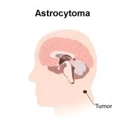
The brain contains many types of cells, including astrocytes, which give neurons the structure and support they need to work well. Neurons form the electrical system that controls brain activity. The most common brain tumor in adults is an astrocytoma, which grows from astrocytes. Doctors diagnose astrocytomas about 15,000 times a year in the US. They occur more often after age 45 and appear more often in men than in women, with a ratio of about 1.3 to 1.
Astrocytoma Defined
Glial cells called astrocytes, found in the brain and spinal cord and shaped like stars, can grow into a type of tumor called an astrocytoma. These uncommon tumors can also spread to other regions of the brain. Older men appear more likely to develop an astrocytoma than children or people of any other age group.
Questions and Answers
What is an Astrocytoma?
An astrocytoma tumor develops from astrocytes, which are a type of glial cell in the brain or spinal cord. Astrocytomas are classified into different grades based on their aggressiveness and potential for growth. Low-grade astrocytomas (grade I and II) are generally slow-growing and less aggressive, while high-grade astrocytomas (grades III and IV) are more aggressive and tend to grow rapidly.
What are the symptoms of an astrocytoma?
The symptoms of an astrocytoma can vary depending on the tumor’s size, location, and grade. Common symptoms may include:
- Headaches: Often persistent and may worsen over time.
- Seizures: Epileptic seizures can occur, especially in low-grade astrocytomas.
- Neurological changes: These may include changes in vision, speech difficulties, memory problems, loss of coordination, weakness, or numbness in certain body parts.
- Cognitive changes: Personality changes, difficulty with concentration, and declining cognitive function may occur as the tumor affects brain function.
How are astrocytomas treated?
The treatment approach for astrocytomas depends on several factors, including the tumor grade, size, location, and the patient’s overall health. The primary treatment options may include:
- Surgery: The surgical removal of the tumor is often the initial treatment for astrocytomas, whenever feasible. A surgeon will remove as much of the tumor as possible without causing damage to critical brain structures.
- Radiation therapy: Doctors will suggest radiation after surgery to target any remaining tumor cells and help prevent recurrence. In some cases, radiation therapy may be used as the primary treatment for inoperable tumors or as palliative care to alleviate symptoms.
- Chemotherapy: Doctors will treat certain types of astrocytomas, particularly high-grade tumors, with chemotherapy. Doctors will administer chemotherapy drugs orally or intravenously to kill or inhibit the growth of cancer cells.
Treatment plans are highly individualized, and a multidisciplinary team of healthcare professionals, including neurosurgeons, oncologists, and radiation therapists, work together to determine the most appropriate course of action for each patient.
Doctors categorize spinal cord Astrocytomas into four classes based on scientific studies in medicine:
Grade I
The surgical excision of certain benign tumors, including pilocytic astrocytoma, can cure them. Although they do not infiltrate other structures, if they get big enough, they can produce symptoms through a mass effect. Most cases of these low-grade astrocytomas appear in youngsters.
Grade II
These are slow-growing and infiltrating spine tumors. Doctors need to regularly monitor them since they can become more aggressive.
Grade III
Also known as Anaplastic Astrocytomas, these aggressive tumors are treated as cancer.
Grade IV
These high-grade astrocytomas are normally malignant and the most aggressive and quickly expanding kind. Higher grade (Grade III) astrocytomas also signify a fast-growing, malignant tumor, while lower grade (Grade I and II) astrocytomas often refer to benign, slow-growing tumors. These cancerous tumors frequently infect neighboring healthy tissue, escalating the harm. Tumors of lower grades can advance to a higher grade.
Additionally, the Location of the Tumors Helps to Categorize Them:
Brainstem gliomas are astrocytomas that develop in this area of the brain. These astrocytomas are situated around the base of the patient’s skull and above the back of the neck, an area where the brainstem aids in controlling vital bodily processes like breathing and heart rate. Although these tumors can be classified as any grade, brainstem gliomas often affect children and young adults and are typically high-grade. Pineal astrocytic tumors are tumors that develop in the region of the pineal gland in the brain. The pea-sized gland responsible for producing the hormone melatonin, which controls the sleep-wake cycle, is located in this area. There are many grades of pineal astrocytic tumors. A few astrocytomas also have oligodendrocytes, a different kind of glial cell. Mixed gliomas or oligoastrocytomas are identified by the names of these tumors.
What Causes an Astrocytoma?
We do not know the exact cause of most astrocytomas. Therapeutic irradiation can lead to the development of astrocytomas. Other environmental exposures, though suspected, do not cause astrocytoma. There is evidence of a hereditary component in some instances, according to several researchers.
Symptoms of Spinal Cord Astrocytoma
The following symptoms may appear, depending on the location of the Spinal Tumor
- chronic back pain
- Emaciation and other sensory modifications
- Inability to control one’s bladder or bowels, Muscle weakness or spasms on one or both sides of the body
- imbalance issues and unstable gait (ataxia)
Doctors identify herniated discs or spinal stenosis as much more likely the source of these symptoms. However, variations can occur that only a skilled expert might spot.
Risk Factors of Spinal Cord Astrocytoma
An astrocyte, or support cell of the nervous system, grows disorderly as a result of a genetic mutation, which results in the development of an astrocytoma. Although a benign or low-grade astrocytoma grows slowly, the effect on the nearby tissues might nonetheless result in symptoms. If more mutations take place, the tumor cells learn how to infiltrate and invade the tissues around them, which results in a malignant transformation. The precise etiology of these alterations is unclear, as it is frequently the case with primary spinal neoplasms. The following identifies known risk factors:
Age
In general, astrocytomas of the spine appear more frequently in children and young people and are less harmful at this age. Adults over the age of 35 usually develop aggressive tumors, which develop into malignancies.
Radiation exposure
A major contributing element to the development of spinal astrocytomas is prior ionizing radiation exposure. Radiation therapy, nuclear pollution, and X-rays provide examples of ionizing radiation. There is no proof linking the development of astrocytomas in the spinal cord to electromagnetic radiation from power lines, microwaves, or mobile phones.
Genetic factors
There is not much proof that hereditary factors play a role in the development of spine astrocytomas.
Diagnosis of Spinal Cord Astrocytoma
Diagnosis begins with a thorough evaluation by a spine specialist. This includes a complete medical history and a neurological exam to identify even subtle defects. When done well, your medical history and physical condition determine which parts of the spine are affected. MRI serves as the test of choice to confirm the diagnosis of astrocytoma and to provide a clear picture of tumor location, size, and shape. Once the tumor is localized and the involvement of surrounding structures is analyzed, the patient usually gets scheduled for a tissue biopsy, either separately or in combination with tumor removal.
A surgical procedure called a biopsy enables the surgeon to take a small sample of the damaged tissue while the patient sleeps in the operating room. A neuropathologist examines the tissue obtained during the biopsy under a microscope in the laboratory. The tumor exam shows if it is benign or malignant. The biopsy also determines the tumor’s grade and cell of origin. Each of these factors guides prognosis and treatment planning.
Treatment of Spinal Cord Astrocytoma
The location, size, grade, age, overall health, and medical history all shape the course of treatment for spinal astrocytomas. If a low-grade astrocytoma shows no signs of growth or spread, careful observation with or without radiation therapy is an option. The safest removal of the tumor without harming the spinal cord is the recommended treatment for large or aggressive tumors. Preserving spinal cord function remains the primary goal. Partial removal can reduce symptoms and slow disease spread, even though it does not cure the condition.
To remove tumors as safely and effectively as possible, a neurosurgeon uses several methods, including lasers and intraoperative ultrasound. Infection, bleeding, spinal fluid leak, weakness, loss of sensation, imbalance, and tumor recurrence are possible post-surgery side effects. Doctors often use chemotherapy and radiation therapy after surgery.
Surgery
Surgery to remove all or as much of the tumor as possible is likely the first step. The exception is when gliomas grow in areas where surgery becomes too risky. Surgery may suffice to cure grade 1 tumors, but it usually does not remove all high-grade tumors.
Radiation Therapy
Radiation is not advised as the first line of treatment for newly diagnosed spinal cord astrocytoma, although it can improve survival when given as adjuvant therapy following surgery. The treatment of recurrent astrocytomas includes radiotherapy as well. The use of radiation therapy (RT) is common:
- To reduce tumors in the spinal cord that do not require surgical removal.
- To reduce the size of the tumor before removal to facilitate surgery.
- To eradicate any remaining tumor cells following an inadequate excision.
A radiation oncologist performs radiation treatment, using a specialized device that accurately directs a beam of high-energy particles (such as X-rays or protons) toward the tumor. Along with other side effects, including nausea, headaches, fatigue, and appetite loss, radiation therapy can also cause red, painful, and irritated skin. Additionally, tingling, weakness, or unbalance may occur.
Chemotherapy
The treatment of glioblastoma and anaplastic astrocytoma with chemotherapy is common. Doctors can either use it before or after radiation. Implanting chemotherapeutic wafers during surgery is a possibility in some circumstances. Although refractory spinal cord Astrocytomas are now the most prevalent use of chemotherapy, their routine usage as adjuvant therapy continues to rise. After surgery, the doctors will discuss having chemotherapy with or without radiation with the parents.
Targeted therapy
Doctors may use a more recent kind of therapy called targeted therapy. This treatment may reduce the size of the tumor. In that it targets certain proteins that promote tumor growth, this differs from chemotherapy in how it functions.
Electric-field therapy
Electric-field treatment employs electrical fields to target tumor cells while sparing healthy cells from damage. Electrodes are placed right on the body to perform the procedure. The system is known as Optune. Following surgery and radiation, it is administered together with chemotherapy. Both those with a recent diagnosis and those whose glioblastoma has returned become eligible for its FDA-approved treatment.
Southwest Scoliosis and Spine Institute specializes in helping patients with Spine Conditions.
If you think you or a loved one might need surgery to correct any kind of complex spine condition, you should contact a surgeon who performs these kinds of complicated and specialized procedures all the time. The Southwest Scoliosis and Spine Institute’s board-certified, fellowship-trained orthopedic surgeons, Richard Hostin, MD, Devesh Ramnath, MD, Ishaq Syed, MD, Shyam Kishan, MD, and Kathryn Wiesman, MD, have performed more than 16,000 successful spine surgeries and are recommended by other doctors all the time. They maintain the highest level of expertise and surgical skills necessary to handle the most complicated cases and achieve successful results.
Significantly, they have also helped more than 100,000 patients get back to living a normal, pain-free life. So, if doctors advised you that nothing can help, please call us at Southwest Scoliosis and Spine Institute, and we have offices in Dallas, Plano, and Frisco, Texas. Finally, with our skills, knowledge, abilities, expertise, and experience, we can offer hope, remove pain, and provide better health. If you need help, call us for an evaluation at 214-556-0555 or visit our contact page today!
____________________
Citation: American Association of Neurological Surgeons – Astrocytoma Tumor
The medical content on this page has been carefully reviewed and approved for accuracy by the Southwest Scoliosis and Spine Institute’s qualified healthcare professionals, including our board-certified physicians and Physician Assistants. Our team ensures that all information reflects the latest evidence-based practices and meets rigorous standards of medical accuracy, with oversight from our expert spine doctors to guarantee reliability for our patients.
We’re here to help STOP THE PAIN
If you are an adult living with scoliosis or have a child with this condition and need a doctor who specializes in orthopedic surgery,
call the Southwest Scoliosis and Spine Institute at 214-556-0555 to make an appointment today.