
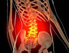
LUMBAR CORD COMPRESSION
Lumbar cord compression occurs when pressure is placed on the spinal cord in the lower back. This can originate from a herniated disc, bone spurs, tumors, or inflammation. Symptoms often include pain, numbness, and weakness in the legs, and can worsen over time. If you experience sudden numbness, severe pain, or loss of bladder or bowel control, seek immediate medical attention by calling The Southwest Scoliosis and Spine Institute. One of our expert Lumbar Cord Compression doctors will quickly see you.


Relief from Lumbar Cord Compression: Stop debilitating leg pain, cramping, and numbness. Our premier spine specialists provide advanced treatments to relieve severe nerve pressure and restore your mobility.
Lumbar Cord Compression:
Lumbar cord compression refers to the narrowing of the spinal canal in the lumbar region. This part of the spine, known as the lumbar spine, consists of five vertebrae located in the lower back, between the ribs and the pelvis. The condition leads to pressure on the spinal cord, which can result from various conditions such as herniated discs, spinal stenosis, tumors, or trauma.
Lumbar spinal stenosis, a common cause of this narrowing, specifically involves the compression of nerves traveling through the lower back into the legs. This compression can cause pain, weakness, numbness, and impaired mobility. Such symptoms significantly impact a person’s quality of life, making early diagnosis and management crucial.
Understanding the precise location and nature of lumbar cord compression helps in identifying effective treatment options and improving patient outcomes.
Lumbar Cord Compression Causes:
Lumbar cord compression can arise from several underlying causes. Herniated discs, where the inner gel-like substance leaks out and presses against the spinal cord, are a common cause.
Spinal stenosis, a narrowing of the spinal canal due to arthritis or bone overgrowth, can also lead to compression. This condition often develops gradually over many years, as several changes occur in the spine.
As we age, our spinal disks lose their sponginess, leading to a reduction in disk height. This change can cause the hardened disks to bulge into the spinal canal. Additionally, bone spurs may develop, and the ligaments within the spine can thicken.
These factors collectively contribute to the narrowing of the central canal. Interestingly, these anatomical changes may not always result in symptoms, highlighting the complex nature of lumbar spinal stenosis.
Understanding these underlying changes is crucial for effectively managing and treating the condition. Additionally, tumors in the spine, spinal injuries, and degenerative conditions can contribute to lumbar cord compression.
Questions and Answers
What is causing my lumbar cord compression, and how severe is it?
Lumbar cord compression can result from various underlying conditions, such as herniated discs, spinal stenosis, or tumors. Your doctor will likely conduct a thorough evaluation, which may include imaging tests like MRI. The MRI will determine the specific cause and severity of the compression. They will then discuss the findings with you and explain how they are contributing to your symptoms.
What treatment options are available for my lumbar cord compression, and which one is best for me?
Treatment for lumbar cord compression depends on the cause of the compression, the severity of symptoms, and your overall health. Your doctor may recommend conservative treatments such as medication, physical therapy, or epidural steroid injections to alleviate symptoms initially. If conservative measures are ineffective or if your symptoms worsen, surgical intervention may be considered. Your doctor will discuss the pros and cons of each treatment option. You and your doctor will make an informed decision based on your circumstances.
What are the potential risks and complications associated with treatment for lumbar cord compression?

Lumbar Cord Compression Symptoms:
Symptoms of lumbar cord compression vary depending on the severity and location of the compression. Common symptoms include:
- Lower back pain that radiates to the legs is often described as “sciatica,” and it can cause pain.
- The weakness or numbness in the legs or feet may extend to the calves and buttocks, impacting daily activities.
- Difficulty walking or maintaining balance, often necessitating frequent short rests due to cramping in the calves while walking.
- Changes in bowel or bladder function may occur rarely but indicate a more severe condition.
- In severe cases, lumbar cord compression can lead to paralysis.
Additionally, symptoms may improve with certain postures, such as bending forward, sitting, or lying down, offering temporary relief. Understanding these symptoms can help in identifying the condition early and seeking appropriate medical advice.
Lumbar Cord Compression Diagnosis:
Non-surgical treatment options for lumbar cord compression aim to alleviate symptoms and improve mobility. This may include medications such as nonsteroidal anti-inflammatory drugs (NSAIDs) to reduce inflammation and pain, physical therapy to strengthen the muscles supporting the spine and improve flexibility, and epidural steroid injections to reduce swelling and alleviate pain.
In addition to these core treatments, several other strategies can enhance recovery and symptom management:
- Posture Management and Exercise: Simple changes in posture and targeted stretching exercises can significantly relieve pressure on the spine. Incorporating a consistent exercise routine can help maintain spinal health and improve overall mobility.
- Weight Management: Maintaining a healthy weight reduces unnecessary stress on the spine, potentially decreasing pain levels and preventing further degeneration.
- Nicotine Cessation: Quitting smoking is crucial as nicotine can impair blood flow and delay healing processes, negatively affecting spinal health.
- Bone-Strengthening Activities: Engaging in activities that promote bone health can provide long-term benefits, ensuring the spine remains resilient and less susceptible to injury.
For most individuals, these approaches can manage pain and enhance quality of life. However, if symptoms persist, consulting a healthcare professional for tailored advice and potential prescription medications might help. Typically, a course of physical therapy lasting four to six weeks is encouraged to achieve optimal results and help patients resume their normal activities.
How is Lumbar Spinal Stenosis Diagnosed?
Diagnosing lumbar spinal stenosis typically involves a thorough assessment by a neurosurgeon. This process includes gathering the patient’s medical history, evaluating symptoms, conducting a physical examination, and analyzing the results of various diagnostic tests.
Common Diagnostic Imaging Tests
To get a comprehensive view of the spine, doctors may order the following imaging studies:
- X-ray: This test uses radiation to capture images of the body’s structures, allowing doctors to examine the alignment and positioning of the spine, bone structure, and joint outlines.
- CT Scan (Computed Tomography): Combining multiple X-ray images, a CT scan provides an in-depth look at the spine’s interior. It offers detailed information about the spinal canal’s size and shape, contents, and surrounding bone structures.
- MRI (Magnetic Resonance Imaging): By using powerful magnets and computer technology, MRI scans generate detailed images of the spinal cord, nerve roots, and adjacent areas. These images help identify issues such as swelling, degenerative changes, or tumors.
- Myelogram: This test involves injecting contrast dye into the space around the spinal cord and nerves. The dye highlights these structures on X-rays and can combine with a CT scan to detect any pressure points affecting the nerves or spinal cord.
By integrating results from these various evaluations, a medical professional can accurately diagnose lumbar spinal stenosis and formulate a suitable treatment plan.
Risk Factors:
Several factors increase the risk of developing lumbar cord compression, including age-related degenerative changes in the spine. In addition, obesity, a sedentary lifestyle, previous spinal injuries, and conditions such as arthritis and osteoporosis can cause this condition. Additionally, occupations or activities that involve repetitive spinal movements or heavy lifting may also increase the risk.
Understanding the Connection Between Degenerative Spondylolisthesis, Degenerative Scoliosis, and Lumbar Spinal Stenosis
Degenerative spondylolisthesis and degenerative scoliosis are two spinal conditions intricately linked with lumbar spinal stenosis. Here’s how they relate:
Degenerative Spondylolisthesis
- Nature of Condition: This occurs when one vertebra slips over the one below it, primarily due to osteoarthritis affecting the facet joints.
- Common Site: The most frequent instance is at the L4-L5 level in the lower back.
- Treatment Approaches: The management strategies—both conservative (non-surgical) and surgical—align closely with those used for lumbar spinal stenosis, focusing on alleviating pressure on the spinal nerves.
Degenerative Scoliosis
- Prevalence and Demographics: Often seen in the lower back, this condition tends to affect individuals over the age of 65.
- Symptoms: The back pain emerges gradually, often linked with physical activities. The spinal curvature is usually mild but can worsen over time.
- Treatment Considerations: Like with spinal stenosis, non-surgical methods are initially employed. However, if these fail, surgical intervention may become necessary to address persistent pain.
Both conditions share a commonality in how they are managed, often requiring similar interventions to relieve symptoms and improve quality of life. Their association with lumbar spinal stenosis primarily revolves around the degenerative changes and the pressure these exert on the spinal canal.
Understanding Lumbar Spinal Stenosis: Development and Demographics
Lumbar spinal stenosis emerges as a condition primarily due to the constriction of the spinal canal in the lower back. This narrowing compresses the nerves that travel from the lower back to the legs. While younger individuals might experience it due to developmental factors, it is predominantly a degenerative issue impacting those aged 60 and over.
Development Process
The development of lumbar spinal stenosis is a gradual process. Over many years, the disks in the spine have lost their natural sponginess, leading to reduced disk height. This can cause the hardened disks to bulge into the spinal canal. Additionally, patients might experience bone spur formation, and the ligaments within the spine can thicken. These changes cumulatively contribute to the narrowing of the spinal canal, though symptoms may vary among individuals.
Who is Affected?
Younger patients can get lumber cord compression due to specific developmental causes; however, the typical demographic comprises older adults. The slow progression over decades often leads to symptoms becoming more pronounced in the senior population.
In summary, lumbar spinal stenosis primarily develops through age-related changes in the spine, making it a condition most commonly seen in those aged 60 and above.
Lumbar Cord Compression Non-Surgical Treatment:
Non-surgical treatment options for lumbar cord compression aim to alleviate symptoms and improve mobility. This may include medications such as nonsteroidal anti-inflammatory drugs (NSAIDs) to reduce inflammation and pain. Physical therapy will also strengthen the muscles supporting the spine and improve flexibility. Meanwhile, epidural steroid injections can reduce swelling and alleviate pain.
In addition to these core treatments, several other strategies can enhance recovery and symptom management:
- Posture Management and Exercise: Simple changes in posture and targeted stretching exercises can significantly relieve pressure on the spine. Incorporating a consistent exercise routine can help maintain spinal health and improve overall mobility.
- Weight Management: Maintaining a healthy weight reduces unnecessary stress on the spine, potentially decreasing pain levels and preventing further degeneration.
- Nicotine Cessation: Quitting smoking is crucial as nicotine can impair blood flow and delay healing processes, negatively affecting spinal health.
- Bone-Strengthening Activities: Engaging in activities that promote bone health can provide long-term benefits, ensuring the spine remains resilient and less susceptible to injury.
For most individuals, these approaches manage pain and enhance quality of life. However, if symptoms persist, consulting a healthcare professional for tailored advice and potential prescription medications should occur. Typically, a course of physical therapy lasting four to six weeks is encouraged to achieve optimal results. It has proven to help patients resume their normal activities.
Lumbar Cord Compression Criteria for Surgical Candidacy in Lumbar Spinal Stenosis
When evaluating if surgery is the right option for lumbar spinal stenosis, several criteria are typically considered:
- Severe Pain Impact: The patient experiences significant back and leg pain that disrupts daily life or diminishes the quality of life.
- Neurological Issues: There is evidence of worsening neurological symptoms, such as weakness in the legs, foot drop, or numbness.
- Bowel and Bladder Dysfunction: The patient encounters issues with normal bowel or bladder control, which may indicate nerve involvement.
- Mobility Challenges: Standing or walking becomes difficult, potentially limiting independence and daily activities.
- Non-Surgical Treatments Ineffective: Prior attempts with medications and physical therapy do not provide sufficient relief.
- Overall Health Status: A healthy patient will undergo surgery safely.
By assessing these factors, healthcare providers can determine if surgical intervention might offer relief and improvement in function that other treatments have not.
Surgical Treatment Details:
In severe cases of lumbar cord compression, where conservative treatments have failed to provide relief or there is progressive neurological deterioration, doctors may recommend surgical intervention. The specific surgical approach depends on various factors, including the underlying cause of the compression, the extent of spinal cord involvement, and the patient’s overall health condition.
Doctors will recommend surgery for lumbar spinal stenosis if:
- Back and leg pain significantly limit normal activity or impair quality of life.
- There is a development of progressive neurological deficits such as leg weakness, foot drop, or numbness in the limb.
- The patient experiences a loss of normal bowel and/or bladder functions.
- There is difficulty standing or walking, impacting daily mobility.
- Medications and physical therapy have proven ineffective in managing symptoms.
- The patient is otherwise in reasonably good health, making them a suitable candidate for surgery.
These criteria help guide the decision-making process, ensuring that surgery is considered when it is most likely to benefit the patient. By addressing these specific factors, healthcare providers can better tailor treatment plans to meet individual needs and improve outcomes.
The following details the surgical treatment options commonly used for severe cases of lumbar cord compression:
Decompression Surgery:
Decompression surgery is aimed at relieving pressure on the spinal cord and nerves by removing or reducing the structures causing compression. This may involve removing portions of herniated discs, trimming overgrown bone (bone spurs), or enlarging the spinal canal to create more space for the spinal cord. The surgical approach for decompression may vary depending on the location and severity of the compression. Common techniques include:
- Laminectomy: Removing the lamina, the bony roof of the spinal canal, to create more space for the nerves. This is often the most common surgery in the lumbar spine, known as a decompressive laminectomy.
- Laminotomy: Surgeons will create a small opening in the lamina, which can be less invasive.
- Foraminotomy: Enlarging the neural foramen, the openings through which spinal nerves exit the spinal canal.
A neurosurgeon may perform a laminectomy with or without additional procedures. These can include fusing vertebrae or removing part of a disk, depending on the patient’s specific needs. The decision for additional procedures depends on the extent of nerve compression and the stability of the spine.
Ultimately, the choice of technique is tailored to relieve pressure effectively while maintaining the structural integrity of the spine, ensuring the best possible outcome for the patient.
What Types of Surgical Procedures Are Available for Treating Lumbar Spinal Stenosis?
There are various surgical approaches to relieve nerve pressure caused by Lumbar spinal stenosis. Here’s a rundown of the key procedures:
- Laminotomy: This procedure involves creating an opening in the lamina, a part of the vertebra, to alleviate nerve root compression without removing the entire structure.
- Foraminotomy: This technique is focused on enlarging the foramen, the passageway where the nerve roots exit the spinal canal, enhancing space for nerve pathways.
- Medial Facetectomy: Part of the facet joint, which can become enlarged or overgrown, is removed to increase space within the spinal canal.
- Anterior Lumbar Interbody Fusion (ALIF): Accessing the spine through the lower abdomen, this method removes a degenerative disk and replaces it with a structural device. This fosters fusion between adjacent vertebrae.
- Posterior Lumbar Interbody Fusion (PLIF): Conducted through the back, this surgery involves removing the disk and posterior spinal bone, retracting nerves, and inserting a supportive device to aid vertebral fusion. This is typically performed on both sides of the spine.
- Transforaminal Lumbar Interbody Fusion (TLIF): Similar to PLIF, TLIF is approached via the back but usually involves one side of the spine. It includes removing the disk and supporting vertebral fusion with a structural device.
- Posterolateral Fusion: Bone grafts are placed along the back and sides of the spine to encourage fusion of the vertebrae.
- Instrumented Fusion: This involves the use of hardware such as screws and rods to stabilize the spine during the fusion process.
Each procedure varies in technique and approach but collectively aims to relieve symptoms and improve stability. Your surgeon can recommend the most appropriate option based on individual diagnoses and conditions.
Surgical Techniques for Treating Lumbar Spinal Stenosis Beyond Decompressive Laminectomy
Decompressive laminectomy is a well-known procedure for lumbar spinal stenosis, but several other surgical techniques can address this condition.
Alternative Surgical Methods
Laminotomy
- Involves creating a small opening in the lamina bone to alleviate nerve root pressure.
Foraminotomy
- Focuses on enlarging the bony exit where the nerve root leaves the spinal canal. Surgeons can perform this procedure in combination with other procedures like laminotomy.
Medial Facetectomy
- This technique involves the removal of part of the facet joint, which may have become overgrown, to increase space within the spinal canal.
Fusion Procedures
Anterior Lumbar Interbody Fusion (ALIF)
- Accesses the spine through the lower abdomen to remove and replace a degenerative disk. A structural implant made from materials like bone or metal is inserted, promoting fusion between adjacent vertebrae.
Posterior Lumbar Interbody Fusion (PLIF)
- Approaches the spine through the back, removing part of the posterior spinal canal bone. The procedure involves nerve retraction to access the disk space, and an implant is placed to facilitate vertebral fusion.
Transforaminal Lumbar Interbody Fusion (TLIF)
- Similar to PLIF, but typically focuses on only one side of the spine. It involves the removal of the posterior bone and insertion of a structural implant after nerve retraction, which supports vertebral fusion.
Posterolateral Fusion
- Involves placing a bone graft on the sides and back of the spine to encourage fusion.
Stability Enhancements
- Instrumented Fusion
- Incorporates hardware such as screws or hooks to stabilize the spine and support the fusion process.
Each of these surgical methods can help a patient’s specific condition, offering various approaches to relieve symptoms and improve spinal stability.
Spinal Fusion:
- Doctors can perform spinal fusion concurrently with decompression surgery, especially in cases where instability of the spine is present. Spinal fusion involves joining two or more vertebrae together using bone grafts, screws, rods, or plates to stabilize the spine and prevent abnormal movement.
- Fusion surgery aims to create a solid union between the vertebrae, which helps maintain spinal alignment and stability. This can help prevent recurrent compression and provide long-term support to the spinal cord and nerves.
Tumor Removal:
- In cases where lumbar cord compression is caused by spinal tumors, surgeons will surgically remove the tumor. The surgical approach for tumor removal depends on factors such as the size, location, and type of tumor.
- Surgeons may perform a complete or partial tumor resection, aiming to remove as much of the tumor as safely possible while preserving neurological function. In some cases, doctors will recommend additional treatments such as radiation therapy or chemotherapy following surgery to target any remaining tumor cells.
Minimally Invasive Surgery:
- Minimally invasive spine surgery techniques may help with decompression and fusion procedures, offering potential benefits such as smaller incisions, reduced tissue damage, shorter hospital stays, and quicker recovery times compared to traditional open surgery.
- Minimally invasive approaches use specialized instruments and imaging guidance to access the spine, allowing surgeons to perform the necessary procedures with precision while minimizing disruption to surrounding tissues.
Postoperative Care and Rehabilitation:
- Following surgery for lumbar cord compression, patients typically require a period of postoperative care and rehabilitation to optimize recovery and regain function. This may include pain management, physical therapy, and gradual return to activities under the guidance of healthcare providers.
- Physical therapy plays a crucial role in strengthening the muscles supporting the spine, improving flexibility, and promoting functional mobility. Patients may also receive education on proper body mechanics and techniques to prevent future spine-related issues.
Patients need to discuss the potential risks, benefits, and expected outcomes of surgical treatment with their healthcare provider, as well as any alternative treatment options available. The decision to undergo surgery for lumbar cord compression should involve the patient and the doctors. It should take into account individual preferences, goals, and overall health status.
Potential Benefits and Risks
When considering surgery for lumbar spinal stenosis, it’s crucial to weigh the potential benefits against the risks involved. A significant number of patients who undergo this surgery report substantial pain relief, which can lead to improved mobility and quality of life. However, it is important to note that surgery might not guarantee relief for everyone.
The risks associated with surgery include not only the inherent challenges of the procedure itself but also those posed by anesthesia. Complications can arise, and outcomes may vary from one individual to another.
By thoroughly discussing these factors with a healthcare provider, patients can make informed decisions that align with their health objectives and circumstances.
Lumbar Cord Compression Complications:
Complications of lumbar cord compression can include persistent pain, weakness, numbness, loss of bladder or bowel control, and paralysis. Without prompt treatment, lumbar cord compression can lead to permanent neurological damage and disability.
Physical Therapy Recommendations:
Physical therapy plays a crucial role in the management of lumbar cord compression, focusing on strengthening the muscles supporting the spine, improving flexibility, and enhancing overall mobility. Therapeutic exercises, manual therapy techniques, and functional training can help alleviate symptoms, improve posture, and prevent further complications.
Prevention Actions:
While some risk factors for lumbar cord compression, such as aging and genetics, cannot be controlled, certain preventive measures can help reduce the risk or delay the onset of symptoms. Also, maintaining a healthy weight, practicing good posture, staying physically active with regular exercise, and avoiding activities that put excessive strain on the spine can help prevent lumbar cord compression.
Related Conditions:
Lumbar cord compression is closely associated with other spinal conditions such as lumbar herniated discs, spinal stenosis, and degenerative disc disease. These conditions often coexist and may contribute to the development or progression of lumbar cord compression.
Living with Lumbar Cord Compression:
Living with lumbar cord compression requires ongoing management and lifestyle adjustments to alleviate symptoms and maintain functionality. Consequently, this may evolve following a treatment plan prescribed by doctors, including medication, physical therapy, and regular monitoring of symptoms. Doctors will recommend assistive devices or mobility aids to enhance mobility and independence.
Long-term Benefits of Treatment and Rehabilitation:
Effective treatment and rehabilitation can significantly improve the long-term outcomes and quality of life for individuals with lumbar cord compression. By addressing the underlying cause of the compression, relieving pressure on the spinal cord, and restoring mobility and function through rehabilitation, individuals can experience reduced pain, improved mobility, and better overall well-being.
Choosing the Southwest Scoliosis and Spine Institute:
When seeking treatment for lumbar cord compression, choosing the right healthcare provider is crucial for optimal outcomes. Finally, the Southwest Scoliosis and Spine Institute, led by renowned spine surgeons Doctors Richard A. Hostin, Devish Ramnath, and Ishaq Syed, offers comprehensive care and state-of-the-art treatments for spinal conditions, including lumbar cord compression. With offices in Dallas, Plano, and Frisco, Texas, the institute provides personalized treatment plans tailored to each patient’s unique needs, ensuring the highest level of care and support throughout the treatment journey.
____________________
Citation: National Institute of Health – Lumbar Cord Compression
The medical content on this page has been carefully reviewed and approved for accuracy by the Southwest Scoliosis and Spine Institute’s qualified healthcare professionals, including our board-certified physicians and Physician Assistants. Our team ensures that all information reflects the latest evidence-based practices and meets rigorous standards of medical accuracy, with oversight from our expert spine doctors to guarantee reliability for our patients.
We’re here to help STOP THE PAIN
If you are an adult living with scoliosis or have a child with this condition and need a doctor who specializes in orthopedic surgery,
call the Southwest Scoliosis and Spine Institute at 214-556-0555 to make an appointment today.