
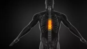
THORACIC CORD COMPRESSION:
Thoracic cord compression occurs when pressure builds up on the spinal cord within the thoracic spine, the middle section of the back. Also, this pressure can pinch the delicate nerves within the cord, disrupting communication between the brain and the body.
Our Thoracic Cord Compression Doctors relieve the pressure or compression on the nerves to relieve pain, burning sensations, or numbness that encircles the chest or abdomen like a belt.


3 percent of individuals with progressive curvature may eventually experience severe problems that can include scoliosis and back pain, spinal problems, and nerve compression causing numbness, weakness, and leg pain.
Thoracic Cord Compression:
 Thoracic cord compression can stem from spine issues like joint swelling, diseases such as rheumatoid arthritis or ankylosing spondylitis. For instance, these conditions cause swelling in spinal joints and nearby tissues. As a result, this leads to spine changes and possible pressure on the spinal cord.
Causes of Thoracic Cord Compression
Thoracic cord compression happens when the spinal cord in the upper back gets pressed, causing nerve issues and trouble with function. Moreover, many conditions can cause this pressure, from spinal wear to injuries or odd spine shapes. Therefore, knowing the causes is key to a correct diagnosis and good treatment. Below are the main causes of thoracic cord compression:
Degenerative Disc Disease (DDD):
Degenerative changes in the intervertebral discs, such as disc herniation, disc bulge, or disc degeneration, can lead to thoracic cord compression. As discs degenerate over time, they may protrude or herniate into the spinal canal, exerting pressure on the spinal cord and nearby nerves.
Spinal Stenosis:
Spinal stenosis refers to the narrowing of the spinal canal, which can compress the spinal cord and nerve roots. In the thoracic region, spinal stenosis may result from hypertrophy of the ligamentum flavum, osteophyte formation, or degenerative changes in the facet joints, reducing the space available for the spinal cord.
Osteoarthritis:
Osteoarthritis of the spine can cause thoracic cord compression by leading to the formation of bone spurs (osteophytes) and thickening of the facet joints. Sometimes, these changes can encroach upon the spinal canal, resulting in compression of the spinal cord and nerve roots.
Traumatic Injuries:
Trauma to the thoracic spine, such as fractures, dislocations, or contusions, can cause direct compression of the spinal cord and surrounding structures. Vehicular accidents, falls, sports injuries, and other traumatic events can result in thoracic cord compression, leading to neurological deficits and functional impairment.
Spinal Tumors:
Tumors originating within the spinal cord (intramedullary tumors), within the spinal canal but outside the spinal cord (intradural-extramedullary tumors), or outside the spinal canal (extradural tumors) can cause compression of the thoracic spinal cord. Surprisingly, spinal tumors may test as benign or malignant and can lead to progressive neurological symptoms if left untreated.
Infections
Infections like spinal abscess, spine tuberculosis, or spinal meningitis can cause swelling in the spinal canal. As a result, this presses on the thoracic spinal cord. In severe cases, abscesses or tissue damage from infections can directly squeeze the spinal cord.
Swelling Disorders
Conditions that cause spine swelling, such as rheumatoid arthritis or ankylosing spondylitis, lead to joint and tissue inflammation. Over time, ongoing swelling may change the spine’s shape, causing deformities and pressure on the thoracic spinal cord.
Birth Defects
Some spine issues present at birth, like spinal dysraphism, tethered cord, or curved spine, can make thoracic cord compression more likely. For example, these birth defects may compress or press the spinal cord, leading to nerve symptoms.
Spine Wear and Curving
Worn-out spine parts paired with side curving (scoliosis) can press on the thoracic spinal cord. As the spine bends oddly, it may push on the cord and nerve roots, causing nerve issues and trouble with function.
Other Causes
Less common causes include blood vessel issues, body chemical problems, immune diseases, or surgery-related scarring. Incidentally, these can press the spinal cord in different ways, needing thorough checks and care.
Conclusion
In summary, thoracic cord compression can come from many causes, like spine wear, injuries, tumors, infections, swelling disorders, birth defects, or other health issues. Therefore, finding the exact cause is key to choosing the right treatment and improving patient results. Finally, a full health check with scans like MRI or CT is vital for a correct diagnosis and to guide treatment options.
Questions and Answers
What causes thoracic cord compression?
Patients commonly inquire about the underlying cause of their symptoms. Patients want to know whether spinal stenosis, herniated discs, tumors, or other spinal abnormalities caused their condition. Answering this question involves a detailed discussion of diagnostic findings from imaging studies. However, it also encompasses neurological evaluations, and medical history to determine the precise cause of cord compression.
What treatment options are available for thoracic cord compression?
Patients often want to understand the available treatment modalities to alleviate their symptoms and improve their quality of life. Doctors will discuss both nonsurgical and surgical treatment options. This includes medications, physical therapy, spinal injections, and surgical interventions like decompression surgery or spinal fusion. Each treatment approach will provide benefits, risks, and considerations, and doctors will thoroughly help patients make informed decisions.
What are the potential long-term effects and prognosis of thoracic cord compression?

Symptoms of Thoracic Cord Compression
Thoracic cord compression occurs when the spinal cord in the thoracic region of the spine is compressed or damaged, leading to a variety of neurological symptoms. The severity of symptoms can vary depending on the underlying cause, the extent of compression, and individual patient factors. Recognizing the symptoms of thoracic cord compression is crucial for timely diagnosis and intervention. Here are the key symptoms associated with this condition:
Back Pain
- A dull, aching pain in the mid-back is a common sign of thoracic cord compression. For example, this pain may stay in the affected spot or spread along the ribs or belly.
Nerve Pain
- Patients may feel sharp or burning pain that spreads along the nerves. Often, this nerve pain moves from the upper back to the chest, belly, or arms.
Numbness and Tingling
- Nerve issues can cause numbness, tingling, or “pins and needles” feelings in the chest, belly, or upper back. Additionally, patients may notice odd sensations or extra sensitivity to touch in these areas.
Muscle Weakness
- Weak muscles in the trunk, belly, or legs can develop due to poor nerve function. As a result, patients may struggle with tasks like walking, standing, or lifting things.
Loss of Coordination
- Problems with balance and coordination can happen when brain-to-muscle signals are disrupted. For instance, patients may feel clumsy, struggle with small tasks, or sense heavy legs.
Bowel or Bladder Issues
- Pressure on the thoracic spinal cord can affect bowel or bladder function. Consequently, patients may face issues like trouble holding urine, urgent need to urinate, leaks, or hard stools.
Movement Problems
- In severe cases, growing weakness or paralysis may occur. Moreover, patients may notice muscle shrinking, low muscle strength, or loss of voluntary movement.
Sensory Loss
- Patients may lose feeling or have less sensitivity to touch, heat, or pain in affected areas. Thus, they might not notice hot or cold or even know they’re hurt.
Spasticity
- High muscle tone and spasticity can come from odd nerve signals. As a result, patients may feel muscle stiffness, unwanted muscle twitches, or trouble moving.
Walking Issues
- Odd walking patterns, like dragging feet, tripping, or balance problems, may show up. These changes often come from weak muscles, sensory loss, or poor coordination.
Conclusion
In summary, thoracic cord compression can cause many symptoms affecting feeling, movement, and body functions. Early spotting of these signs is key to quick diagnosis and starting the right treatment to avoid nerve issues and improve patient results. Therefore, anyone with these symptoms should seek a doctor’s care for further care and treatment.
Methods of Diagnosing Thoracic Cord Compression
Diagnosing thoracic cord compression involves a combination of clinical evaluation, imaging studies, and diagnostic tests to assess spinal cord function and identify the underlying cause of compression. Timely and accurate diagnosis is crucial for implementing appropriate treatment strategies and preventing neurological complications. Here are the key methods used in diagnosing thoracic cord compression:
Medical History and Physical Examination:
- A thorough medical history helps identify risk factors, previous injuries, and symptoms suggestive of thoracic cord compression.
- During a physical exam, the healthcare provider assesses muscle strength, reflexes, sensation, and coordination to detect problems of spinal cord dysfunction.
Imaging Studies:
- MRI (Magnetic Resonance Imaging): MRI is the primary imaging modality used to visualize the spinal cord, nerve roots, and surrounding structures with high resolution.
- MRI can identify the location, extent, and severity of spinal cord compression, as well as detect abnormalities such as herniated discs, tumors, or spinal stenosis.
- CT (Computed Tomography) Scan: Doctors may authorize CT scans to provide detailed images of the bony structures of the spine and assess for fractures, bone spurs, or other abnormalities that may contribute to cord compression.
- X-rays: Doctors may authorize X-rays to evaluate the alignment of the spine and identify fractures, dislocations, or degenerative changes.
Electrodiagnostic Testing:
- Electromyography (EMG) and Nerve Conduction Studies (NCS): EMG and NCS are used to assess the electrical activity of muscles and nerves, respectively.
- These tests can help determine the extent of nerve damage, identify the location of nerve compression or injury, and differentiate between nerve root and spinal cord involvement.
CSF Analysis:
- In some cases, doctors will perform a lumbar puncture (spinal tap) to collect cerebrospinal fluid (CSF) for analysis.
- CSF analysis may help identify inflammatory or infectious causes of spinal cord compression, such as meningitis or spinal cord tumors.
Neurological Evaluation:
- Neurological assessments, including sensory testing, reflex testing, and assessment of muscle strength and coordination, help evaluate the severity and progression of spinal cord dysfunction.
- Basically, doctors will perform neurological examinations periodically to monitor changes in neurological status and response to treatment.
Specialized Tests:
- Depending on the suspected cause of thoracic cord compression, doctors may request additional specialized tests.
- These may include genetic testing for hereditary conditions, angiography for vascular abnormalities, or biopsy for suspected tumors or inflammatory disorders.
In conclusion, the diagnosis of thoracic cord compression relies on a comprehensive approach involving clinical evaluation, imaging studies, and specialized tests. Collaboration between healthcare providers, neurologists, and radiologists is essential to accurately diagnose the condition and develop an individualized treatment plan for optimal patient outcomes. Early diagnosis and intervention are critical to prevent neurological deficits and improve long-term prognosis for patients with thoracic cord compression.
Nonsurgical Treatment
Nonsurgical treatment options for thoracic cord compression may include medications to manage pain and inflammation, physical therapy to improve strength and mobility, and lifestyle modifications to reduce strain on the spine. In some cases, doctors may recommend bracing or orthotic devices to provide additional support to the spine.
Surgical Treatment
Surgical treatment options for thoracic cord compression are typically considered when conservative measures fail to alleviate symptoms or when there is evidence of progressive neurological deficits. The specific surgical approach depends on factors such as the underlying cause of compression, the extent of spinal cord involvement, and the patient’s overall health. Here are the surgical options commonly used for thoracic cord compression:
Decompressive Surgery:
- Decompressive surgery aims to alleviate pressure on the spinal cord by removing or reshaping the structures causing compression, such as herniated discs, bone spurs, or tumors.
- Procedures may include laminectomy, laminoplasty, or discectomy, depending on the location and nature of the compression.
- Laminectomy involves removing a portion of the vertebral bone (lamina) to create more space for the spinal cord.
- Laminoplasty is a technique used to expand the spinal canal by reshaping the lamina without removing it entirely.
- Discectomy involves removing part or all of a herniated disc that is compressing the spinal cord.
Spinal Fusion:
- Surgeons may perform spinal fusion in conjunction with decompressive surgery to stabilize the spine and prevent future instability or deformity.
- During spinal fusion, bone grafts, metal implants, or bone substitutes are used to fuse adjacent vertebrae, limiting movement at the affected segment.
- Surgeons may perform spinal fusion anteriorly (from the front), posteriorly (from the back), or both, depending on the surgical approach and the extent of spinal instability.
Tumor Resection:
- In cases where spinal tumors cause thoracic cord compression, surgical resection of the tumor may be necessary.
- The goal of tumor resection is to remove as much of the tumor as possible while preserving neurological function.
- Depending on the tumor’s location and characteristics, surgeons may perform surgery using minimally invasive spine surgery or open surgical approaches.
Spinal Cord Detethering:
- Tethered spinal cord syndrome occurs when the spinal cord is abnormally attached to surrounding tissues, causing tension and compression.
- Surgical detethering involves releasing the adhesions and restoring the spinal cord’s normal mobility and function.
- This procedure may be indicated for patients with conditions such as tethered cord syndrome or spinal dysraphism.
Vertebroplasty or Kyphoplasty:
- Vertebroplasty and kyphoplasty are minimally invasive procedures used to treat vertebral compression fractures caused by osteoporosis or trauma.
- During vertebroplasty, bone cement is injected into the fractured vertebra to stabilize it and relieve pain.
- Kyphoplasty involves inserting a balloon-like device into the fractured vertebra to restore height before injecting bone cement.
Dynamic Stabilization:
- Dynamic stabilization techniques involve implanting devices such as pedicle screws, rods, or interspinous spacers to provide stability while preserving some degree of spinal mobility.
- These procedures may be considered in select cases to address thoracic cord compression while allowing for physiological movement of the spine.
Each surgical option for thoracic cord compression carries its risks, benefits, and considerations. The choice of procedure depends on factors such as the underlying cause of compression, the patient’s overall health, and the surgeon’s expertise and preferences. Patients need to discuss the potential risks and benefits of each surgical option with their healthcare provider to make informed decisions about their treatment.
Benefits of Surgical Treatment
The benefits of surgical treatment for thoracic cord compression include the relief of neurological symptoms. In addition, the benefits will preserve and restore neurological functions, and prevent further spinal cord damage. Surgery can also improve the overall quality of life and functional outcomes for patients with this condition.
Recovery Period and Rehabilitation
The recovery period following surgery for thoracic cord compression can vary depending on the extent of the procedure and individual factors such as overall health and fitness level. Rehabilitation may include physical therapy, occupational therapy, and other modalities to promote recovery, improve strength and mobility, and prevent complications.
Reasons to Choose The Southwest Scoliosis and Spine Institute:
Finally, patients with Thoracic Cord Compression can benefit from the expertise of the renowned spine surgeons at The Southwest Scoliosis and Spine Institute. Led by Doctors Richard Hostin, MD, Devesh Ramnath, MD, Ishaq Syed, MD, Shyam Kishan, MD, and Kathryn Wiesman, MD, the institute offers state-of-the-art diagnostic and treatment options for complex spinal conditions. With offices in Dallas, Plano, and Frisco, Texas, the institute provides personalized care and comprehensive support to help patients achieve optimal outcomes and improve their quality of life.
____________________
Citation: ScienceDirect.com – Thoracic Cord Compression
The medical content on this page has been carefully reviewed and approved for accuracy by the Southwest Scoliosis and Spine Institute’s qualified healthcare professionals, including our board-certified physicians and Physician Assistants. Our team ensures that all information reflects the latest evidence-based practices and meets rigorous standards of medical accuracy, with oversight from our expert spine doctors to guarantee reliability for our patients.
We’re here to help STOP THE PAIN
If you are an adult living with scoliosis or have a child with this condition and need a doctor who specializes in orthopedic surgery,
call the Southwest Scoliosis and Spine Institute at 214-556-0555 to make an appointment today.