
THORACIC DISC EXTRUSION:
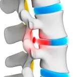
Thoracic disc extrusion, also known as a herniated thoracic disc, occurs when the soft inner core (nucleus pulposus) of an intervertebral disc in the thoracic spine (mid-back region) pushes through a tear in the tough outer shell (annulus fibrosus). This extruded disc material can press on the spinal cord or nerve roots in the area, causing pain, numbness, and weakness.
Unlike the lower back (lumbar spine), where disc herniations are more frequent, thoracic disc extrusions are rare. They can be challenging to diagnose due to the smaller size of the thoracic area spinal canal — even by Thoracic Disc Extrusion Doctors and specialists. Also, the presence of ribs limits some diagnostic tests.


Elite Care for Mid-Back Disc Ruptures: Trust the leading neurosurgical team at Southwest Scoliosis and Spine Institute to execute sophisticated, micro-endoscopic decompressions for complex thoracic disc extrusions.
Thoracic Disc Extrusion
 The term thoracic disc extrusion refers to a condition characterized by the rupture of the inner gel-like substance of a spinal disc through the tough outer layer, leading to compression of the spinal cord or nerve roots in the thoracic region of the spine. Thoracic Disc Extrusion is relatively rare as compared to disc extrusions in the cervical or lumbar spine. Also, thoracic disc extrusion can cause significant pain, neurological symptoms, and functional impairment. It is often associated with spinal inflammatory disorders such as discitis, spondylodiscitis, and ankylosing spondylitis.
The Causes of Thoracic Disc Extrusion
Thoracic disc extrusion occurs when the inner gel-like substance of a spinal disc ruptures through the tough its outer layer. This leads to compression of the spinal cord or nerve roots in the thoracic region of the spine. Several factors can contribute to the development of thoracic disc extrusion, including:
- Age-related degeneration: As individuals age, the spinal discs undergo wear and tear, causing them to lose their elasticity and resilience. This degeneration can weaken the outer layer of the disc, making it more susceptible to herniation or extrusion.
- Traumatic injury: Trauma to the thoracic spine, such as a sudden impact or fall, can exert excessive force on the spinal discs. Trauma can lead to displacement or rupture. Traumatic injuries may occur in accidents, sports-related activities, or falls.
- Underlying spinal conditions: Certain spinal conditions, such as disc herniation, spinal stenosis, or degenerative disc disease, can predispose individuals to thoracic disc extrusion. These conditions may alter the structure and integrity of the spinal discs, making them more prone to herniation or extrusion.
- Repetitive strain or overuse: Activities that involve repetitive bending, lifting, or twisting motions can place stress on the spinal discs. This will increase the risk of disc degeneration and extrusion over time. Occupations or hobbies that require repetitive spinal movements may contribute to the development of thoracic disc extrusion.
- Genetic predisposition: In some cases, a genetic component to the development of thoracic disc extrusion. Certain genetic factors or hereditary conditions may influence the structure and composition of the spinal discs. When this occurs, it increases the likelihood of disc degeneration and extrusion.
Overall, thoracic disc extrusion is a multifactorial condition influenced by the above conditions. Understanding these potential causes is essential for accurate diagnosis and appropriate management of thoracic disc extrusion.
Questions and Answers
What causes thoracic disc extrusion?
Thoracic disc extrusion occurs when the gel-like material inside a spinal disc protrudes through the outer layer, typically due to disc degeneration, trauma, or repetitive stress. Causes may include age-related wear and tear, injury from lifting or twisting, or underlying spinal conditions such as disc herniation or degenerative disc disease.
What are the symptoms of thoracic disc extrusion?
Symptoms of thoracic disc extrusion can vary depending on the severity and location of the extruded disc material. Common symptoms may include mid-back pain, radiating pain or numbness in the chest or abdomen, weakness or paralysis in the legs, difficulty walking or maintaining balance, and changes in bowel or bladder function. Severe cases may lead to spinal cord compression and neurological deficits.
What treatment options are available for thoracic disc extrusion?

Understanding the Types of Thoracic Outlet Syndrome
Thoracic Outlet Syndrome (TOS) can manifest in three primary forms, each affecting the body in distinct ways due to compression issues within the thoracic outlet area.
Neurogenic Thoracic Outlet Syndrome (nTOS)
This is the most prevalent type, accounting for over 90% of cases. Neurogenic TOS occurs when there’s compression of the brachial plexus, the nerve bundle that carries signals from the spinal cord to the shoulder, arm, and hand. Symptoms often include pain, weakness, or numbness in these areas.
Venous Thoracic Outlet Syndrome (vTOS)
Venous TOS makes up about 5% of cases and involves the compression of a vein. This type results in symptoms such as swelling, discoloration, and possibly pain due to the restricted blood flow. In severe cases, it can lead to upper-body thrombosis.
Arterial Thoracic Outlet Syndrome (aTOS)
This is the rarest form, representing approximately 1% of cases. Arterial TOS is caused by the compression of an artery, which can lead to a lesser blood supply to the arm and hand. This may result in severe pain, cold sensitivity, and weakened muscles.
Vascular Thoracic Outlet Syndrome
Both venous and arterial types are collectively referred to as Vascular Thoracic Outlet Syndrome, highlighting their impact on blood vessels rather than nerves. Understanding these subtypes helps in diagnosing and tailoring a treatment approach for those afflicted.
What Are the Treatment Options for Neurogenic Thoracic Outlet Syndrome?
Neurogenic Thoracic Outlet Syndrome (TOS) can significantly impact daily life, but there are several treatment strategies available to alleviate symptoms.
- Physical Therapy: Often the first line of defense, a tailored physical therapy program focuses on relieving symptoms through exercises that improve posture and reduce pressure on the nerves.
- Botulinum Toxin Injections: When physical therapy does not fully resolve the symptoms, injections of this drug can provide relief by relaxing specific muscles.
- Surgical Intervention: If symptoms persist, surgery might be the next step. This could involve procedures such as releasing the anterior and middle scalene muscles or removing the first rib to ease nerve compression.
Considerations for Recurrence
Despite treatment, neurogenic TOS might re-emerge months or even years later. This can occur due to scar tissue formation at the surgical site or initial misdiagnosis. Ongoing monitoring and follow-up with your doctor is crucial to address any recurrence of symptoms gracefully.
Understanding the Differences Between Neurogenic and Vascular Thoracic Outlet Syndrome
Thoracic Outlet Syndrome (TOS) manifests in various forms, primarily distinguished by the affected body structures: nerves, veins, or arteries. Each variant of TOS presents distinct features and prevalence.
Neurogenic Thoracic Outlet Syndrome
- Definition: This form involves the compression of the brachial plexus, the network of nerves extending from the neck to the arm.
- Prevalence: It is the most common type, accounting for over 90% of all TOS cases.
- Symptoms: Often include pain, tingling, and weakness in the neck, shoulder, and arm.
Vascular Thoracic Outlet Syndrome
Venous TOS:
- Details: Occurs when a vein is compressed.
- Prevalence: Represents around 5% of TOS cases.
- Symptoms: Can lead to upper extremity swelling and pain.
Arterial TOS:
- Details: Involves compression of an artery.
- Prevalence: The rarest form, constituting about 1% of cases.
- Symptoms: May result in coldness, paleness, or weak pulses in the arm.
Together, venous and arterial TOS are often grouped under Vascular Thoracic Outlet Syndrome because they both affect blood vessels. The key distinction between neurogenic and vascular TOS lies in the specific structures compressed—nerves versus blood vessels—and their symptoms and frequency. Understanding these differences is crucial for appropriate diagnosis and treatment.
Symptoms
Thoracic disc extrusion occurs when the gel-like substance of a spinal disc protrudes through a tear in the disc’s outer layer, potentially compressing the spinal cord or nerve roots in the thoracic region of the spine. The symptoms of thoracic disc extrusion can vary depending on the severity of the compression and the specific nerves affected. Common symptoms may include:
Thoracic back pain:
- Pain in the middle or upper back is a common symptom of thoracic disc extrusion. The pain only occurs in the affected area of the spine and may worsen with movements such as bending, twisting, or lifting.
Radiating pain:
- In some cases, thoracic disc extrusion can cause pain that radiates along the path of the affected nerves. This may result in pain that travels around the rib cage, chest, or abdomen. The pain may appear as sharp, stabbing, or burning in nature.
Numbness or tingling:
- Compression of the spinal cord or nerve roots in the thoracic spine can lead to sensory changes, such as numbness, tingling, or a pins-and-needles sensation. These sensations may occur in the back, chest, abdomen, or other areas supplied by the affected nerves.
Muscle weakness:
- Thoracic disc extrusion can cause weakness in the muscles of the back, abdomen, or lower extremities, depending on the location and severity of the compression. Muscle weakness may affect mobility and coordination, leading to problems with walking or performing daily activities.
Bowel or bladder dysfunction:
- In severe cases of thoracic disc extrusion, compression of the spinal cord may disrupt nerve signals that control bowel and bladder function. This can lead to symptoms such as urinary or bowel problems.
Difficulty breathing:
- Rarely, a thoracic disc extrusion may compress nerves that supply the muscles involved in breathing, leading to respiratory difficulties. This may manifest as shortness of breath, chest tightness, or difficulty taking deep breaths.
It’s important to note that the symptoms of thoracic disc extrusion can vary from person to person, and some individuals may experience only mild or intermittent symptoms. However, persistent or worsening symptoms should prompt evaluation by a doctor to determine the underlying cause.
Diagnosis
Diagnosing thoracic disc extrusion typically involves a combination of clinical evaluation, imaging studies, and sometimes additional diagnostic tests. Here’s an overview of the process and tools used by doctors to diagnose this condition:
Medical History and Physical Examination:
- The diagnostic process often begins with a thorough medical history and physical examination. The doctor will inquire about the patient’s symptoms, medical history, and any recent injuries or trauma to the spine.
- During the physical exam, the doctor will assess the patient’s range of motion, muscle strength, sensation, and reflexes. Doctors may order specific tests to evaluate for signs of spinal cord or nerve compression.
Imaging Studies:
Imaging studies are crucial for confirming the presence of thoracic disc extrusion and assessing its severity. The most common imaging modalities used include:
- MRI (Magnetic Resonance Imaging): MRI is the preferred imaging modality for evaluating the spine and detecting disc herniation. It provides detailed images of the spinal cord, nerve roots, and intervertebral discs, allowing the doctor to visualize the location and extent of the disc extrusion.
- CT (Computed Tomography) Scan: In some cases, the doctor will order a CT scan to obtain additional info about the bony structures of the spine, especially if surgery is being considered.
- Electrodiagnostic Testing (Optional):
- In certain situations, doctors will order electrodiagnostic tests or nerve conduction studies (NCS) to assess nerve function and determine the extent of nerve damage. These tests can help doctors know the difference between nerve compression due to disc extrusion and other neurological conditions.
Diagnostic Injections:
- In some cases, doctors will use diagnostic injections to confirm the source of pain and assess the response to treatment. These injections involve injecting a medication into the area surrounding the affected nerve roots or spinal cord.
Clinical Correlation:
Once the diagnostic tests are completed, the doctor will correlate the findings with the patient’s clinical presentation to make an accurate diagnosis of thoracic disc extrusion. This includes considering the location and size of the extruded disc, the presence of spinal cord or nerve compression, and the severity of symptoms.
Overall, a comprehensive approach combining medical history, physical examination, and imaging studies is essential for diagnosing thoracic disc extrusion and determining the most appropriate treatment plan for each patient.
How is Thoracic Outlet Syndrome Diagnosed?
Diagnosing thoracic outlet syndrome (TOS) can be challenging due to its often vague symptoms, which can mimic other conditions. Proper diagnosis requires an approach to differentiate between the various types of TOS and exclude similar conditions.
Initial Evaluation Steps
- Medical History and Symptom Review: The first step involves gathering a detailed medical history and understanding the patient’s symptoms. This helps identify potential patterns indicative of TOS.
- Physical Exam: Healthcare providers perform specific physical maneuvers designed to reproduce symptoms, providing clues to the presence of TOS.
- Exclusion of Other Conditions: Evaluating the patient’s symptoms to rule out other nerve-related disorders—such as carpal tunnel syndrome, cubital tunnel syndrome, or cervical spine issues—is crucial. This may involve nerve conduction studies or cervical spine MRI to ensure these conditions are not mistaken for TOS.
Diagnostic Tests
- Duplex Ultrasound: This test is utilized to detect any narrowing or blockage in blood vessels, which could suggest vascular TOS.
- Chest X-ray: This imaging helps identify problems like a cervical rib or an unusually formed first rib, which might cause symptoms.
Specific Tests for Neurogenic TOS
- Brachial Plexus Block: In suspected neurogenic TOS cases, a local anesthetic is injected into the neck’s scalene muscles. If this procedure alleviates symptoms, the likelihood of neurogenic TOS is increased.
By following this diagnostic strategy, healthcare providers can accurately diagnose thoracic outlet syndrome and separate it from other disorders with overlapping symptoms.
Treatment Options for Thoracic Disc Extrusion
Treatment typically depends on the severity of symptoms, the extent of spinal cord or nerve compression, and the patient’s overall health. Here’s an overview of nonsurgical and surgical options:
Nonsurgical Treatment:
Medications: Doctors will prescribe nonsteroidal drugs (NSAIDs) to reduce pain and inflammation associated with thoracic disc extrusion.
Physical Therapy: A structured physical therapy program can help improve flexibility, strengthen the muscles supporting the spine, and alleviate symptoms. Therapeutic exercises, manual therapy, and modalities like heat or ice may be included.
Activity Modification: Avoiding activities that cause symptoms, such as heavy lifting or prolonged sitting, can help prevent further aggravation of the condition. Epidural Steroid Injections: In cases where conservative measures fail to provide relief, epidural steroid injections will deliver medication directly into the affected area. The injections will temporarily reduce pain and inflammation.
How Botulinum Toxin Injections Aid in Treating Thoracic Outlet Syndrome
Thoracic outlet syndrome (TOS) can be a condition to manage, particularly when traditional therapies don’t bring sufficient relief. For some patients experiencing persistent symptoms, other medicines may offer an effective alternative.
What are Botulinum Toxin Injections?
Botulinum toxin, known for its muscle-relaxing properties, can be injected directly into the affected area. This works by blocking nerve signals that cause muscles to contract excessively, thereby reducing pressure on the nerves and blood vessels in the thoracic outlet.
Why Consider Botulinum Toxin for TOS?
- Muscle Relaxation: It helps relax overactive muscles, lessening muscle compression on nerves and vessels.
- Symptom Relief: Many patients experience a significant reduction in pain and discomfort.
- Targeted Approach: The injection is precise, targeting areas where physical therapy and other treatments might not reach.
Treatment Process
- Evaluation: A thorough assessment by a doctor will ensure the patient is a suitable candidate for the injections.
- Injection: Administered by a specialist, often under imaging guidance to ensure precision.
- Follow-Up: Post-treatment evaluations help monitor healing and adjust treatment plans.
Potential Benefits
Patients who use botulinum toxin often notice an improvement in mobility and a reduction in pain levels, making daily activities more manageable. While it may not be the first line of treatment, it serves as a valuable option when other therapies fall short.
By employing botulinum toxin injections, doctors can directly address muscle-related issues contributing to the disorder, providing patients with a tailored path toward relief.
Why Might Thoracic Outlet Syndrome Recur After Treatment?
Thoracic outlet syndrome (TOS) can, unfortunately, make a comeback even after seemingly successful treatment. There are a couple of reasons why this might happen:
- Scar Tissue Development: After surgery, scar tissue can form around the treated area. This scar tissue can put pressure on nerves or blood vessels, potentially causing symptoms to reappear.
- Misdiagnosis of the Condition: In some cases, the initial diagnosis might not have accurately pinpointed the root cause of symptoms. If the underlying issue wasn’t fully addressed, symptoms of TOS can resurface over time.
It’s crucial to monitor post-treatment symptoms and consult with healthcare providers if any concerns arise. Understanding these factors can help manage expectations and guide post-treatment care.
Surgical Treatment:
- Decompressive Surgery: Doctors will recommend surgical intervention if the patient experiences severe or progressive neurological deficits, such as weakness, numbness, or difficulty walking, due to spinal cord or nerve compression.
- Discectomy: During a discectomy, the surgeon removes the portion of the herniated or extruded disc that is compressing the spinal cord or nerve roots. This procedure aims to relieve pressure on the affected structures and alleviate symptoms.
- Laminectomy: In some cases, a surgeon will perform a laminectomy to remove a portion of the vertebral bone (lamina) to create more space within the spinal canal and relieve pressure on the spinal cord.
- Spinal Fusion: In instances where there is significant instability or deformity of the spine, doctors will recommend spinal fusion to stabilize the affected segment and prevent further progression of the condition. During fusion surgery, the surgeon uses bone grafts and implants to fuse two or more adjacent vertebrae, promoting bone growth and stability.
- Minimally Invasive Techniques: Surgeons will use minimally invasive spine surgery, such as microdiscectomy or endoscopic discectomy, for select patients with thoracic disc extrusion. These techniques involve smaller incisions, reduced muscle damage, and faster recovery compared to traditional open surgery.
The choice between nonsurgical and surgical treatment options for thoracic disc extrusion should be made in consultation with a spine specialist and tailored to each patient’s individual needs, preferences, and overall health status.
What are the Treatment Options for Arterial Thoracic Outlet Syndrome?
Arterial Thoracic Outlet Syndrome (TOS) often necessitates surgical intervention. Here’s a breakdown of the treatment options available:
Surgical Intervention
Surgery is frequently the primary approach for addressing arterial TOS. It typically involves removing specific anatomical structures such as:
- Scalene Muscles: These muscles in the neck may be excised to alleviate pressure.
- Cervical Rib: If present, this extra rib may be removed to improve blood flow.
- First Rib: This may also be removed to relieve compression on arteries and nerves.
Medical Management
In conjunction with or as an alternative to surgery, doctors may suggest the following treatments:
- Blood Thinners: These medications are used to dissolve blood clots that might have formed due to restricted blood flow.
Vascular Procedures
For more serious cases, doctors may implement additional procedures:
- Arterial Reconstruction or Replacement: If the affected artery shows signs of an aneurysm or contains a clot, reconstructive surgery or complete artery replacement might be required to restore normal circulation.
By tailoring these treatment strategies to the patient’s specific condition, healthcare professionals aim to minimize symptoms and improve quality of life.
What Are the Treatment Options for Venous Thoracic Outlet Syndrome?
When it comes to managing venous Thoracic Outlet Syndrome (TOS), surgery is often the cornerstone of treatment. The goal is to relieve pressure on the vein, typically requiring the removal of certain anatomical structures like the scalene and subclavius muscles, as well as the first rib.
However, doctors must address the damaged vein to prevent complications from blood clots that can develop on its inner surface. Here are the primary treatment options:
- Blood Thinners: Medications are commonly prescribed to manage and reduce blood clots.
- Thrombolysis: This procedure aims to dissolve any existing clots in the vein and is generally performed before the main surgical intervention for TOS.
- Post-Surgery Monitoring: After rib resection surgery, doctors may suggest a venogram a few weeks later. This imaging test assesses any lingering damage to the vein. If necessary, doctors will conduct a balloon angioplasty, which involves inflating a small balloon inside the vein to widen any narrowed areas.
These treatments are designed to alleviate symptoms and restore normal blood flow, aiming for a comprehensive approach to venous TOS.
The Benefits of Surgery
Surgery for thoracic disc extrusion aims to alleviate symptoms, reduce spinal cord or nerve compression, and improve the overall quality of life for patients. Here are some potential benefits of surgical intervention for thoracic disc extrusion:
Relief of Symptoms:
- Surgery can effectively alleviate symptoms associated with thoracic disc extrusion, such as pain, numbness, tingling, weakness, and difficulty walking. By removing the herniated or extruded disc material that is compressing the spinal cord or nerve roots, surgery can provide immediate relief of neurological deficits and improve function.
Prevention of Neurological Damage:
- Thoracic disc extrusion can lead to progressive neurological deficits if left untreated, potentially resulting in permanent nerve damage and loss of function. Surgical decompression of the spinal cord or nerve roots can help prevent further neurological deterioration and may even facilitate recovery of function in some cases.
Improved Mobility and Function:
- By alleviating spinal cord or nerve compression, surgery can improve mobility, strength, and overall function for patients with thoracic disc extrusion. This may enable patients to resume activities of daily living, return to work, and engage again in recreational pursuits.
Reduced Pain and Discomfort:
- Surgery can significantly reduce or eliminate pain and discomfort caused by thoracic disc extrusion. By addressing the underlying cause of pain and inflammation, surgery can provide long-term pain relief.
Stabilization of the Spine:
- In cases where there is spinal instability or deformity due to disc extrusion, spinal fusion surgery may occur. Spinal fusion can prevent further progression, reduce the risk of recurrent disc herniation, and promote long-term spinal stability.
Faster Recovery and Return to Activity:
- With advances in surgical techniques and technology, many patients experience faster recovery and shorter hospital stays following surgery. Minimally invasive approaches, in particular, can result in smaller incisions, reduced muscle damage, and a quicker return to normal activities.
Improved Quality of Life:
- Ultimately, surgical intervention for thoracic disc extrusion aims to improve the overall quality of life for patients. The goal is to relieve symptoms, restore function, and prevent long-term neurological complications. By addressing the underlying pathology, surgery can help patients regain independence, mobility, and overall well-being.
It’s important to note that the decision to undergo surgery for thoracic disc extrusion should include consultation with a spine specialist. Both should discuss weighing the potential benefits against the risks and considering each patient’s circumstances and treatment goals.
The Recovery
Recovery from surgery for thoracic disc extrusion can vary depending on various factors. These include the surgical procedure performed, the extent of the condition, the patient’s overall health, and adherence to postoperative instructions. Here’s a general overview of the recovery period:
Immediate Postoperative Period (Hospital Stay):
- After surgery, patients typically spend a few days in the hospital for close monitoring and pain management.
- During this time, doctors will assess the patient’s neurological functions, monitor for complications, and provide instructions for postoperative care.
Initial Recovery at Home (First Few Weeks):
- Once discharged from the hospital, patients will need to rest and gradually increase their activity level as tolerated.
- Doctors may prescribe pain management medications to alleviate discomfort during the initial recovery period.
- Patients are advised to avoid strenuous activities, heavy lifting, and bending forward to prevent strain on the surgical site.
- Patients may begin physical therapy or rehabilitation to help restore strength, flexibility, and mobility gradually.
Mid-Term Recovery (Several Weeks to Months):
- Over the next several weeks to months, patients typically experience a gradual improvement in symptoms and function.
- Follow-up appointments with the surgeon will monitor progress and adjust the treatment plan as needed.
- Physical therapy or rehabilitation focuses on strengthening the muscles supporting the spine, improving posture, and enhancing overall mobility.
- Patients may gradually resume activities of daily living, including light household chores, walking, and gentle stretching exercises.
Long-Term Recovery (Months to Years):
- Full recovery from surgery for thoracic disc extrusion may take several months to a year or longer, depending on individual factors.
- While most patients experience significant symptom relief and functional improvement, some residual symptoms or limitations may persist.
- Patients need to continue following their surgeon’s recommendations. Especially for activity modification, rehabilitation exercises, and any prescribed medications.
- Regular follow-up visits with the surgeon will monitor long-term outcomes and address any ongoing concerns.
Overall, the recovery timeline following surgery for thoracic disc extrusion can vary widely among patients. Individuals must communicate with their doctors, adhere to postoperative guidelines, and actively participate in their rehabilitation program.
Rehabilitation
Rehabilitation following surgery for thoracic disc extrusion plays a vital role in promoting healing, restoring function, and preventing future complications. The rehabilitation program may vary depending on the individual’s condition, the surgical procedure performed, and the patient’s overall health status. However, here are some common components of rehabilitation for patients undergoing surgery for thoracic disc extrusion:
Early Mobilization and Range of Motion Exercises:
- In the initial stages of rehabilitation, patients are encouraged to begin gentle mobilization exercises to prevent stiffness and promote blood circulation.
- Range of motion exercises, including gentle stretching of the spine and surrounding muscles, maintain flexibility and prevent muscle tightness.
Core Strengthening Exercises:
- Strengthening the muscles that support the spine, particularly the core muscles, is essential for stability and proper spinal alignment.
- Physical therapists may prescribe exercises targeting the abdominal muscles, lower back muscles, and pelvic floor muscles to improve core strength and stability.
Postural Training:
- Proper posture is crucial for reducing strain on the spine and preventing recurrent injuries.
- Patients may receive instruction on ergonomic principles and posture correction techniques to maintain a neutral spine position during daily activities.
Cardiovascular Conditioning:
- Aerobic exercise, such as walking, stationary cycling, or swimming, can help improve cardiovascular fitness and overall endurance.
- Patients may gradually incorporate cardiovascular activities into their rehabilitation program under the guidance of a healthcare professional.
Functional Training:
- Functional exercises aim to simulate activities of daily living and improve functional capacity.
- Patients may perform tasks such as bending, lifting, reaching, and squatting under the supervision of a therapist to enhance functional independence and confidence.
Pain Management Techniques:
- Modalities such as ice or heat therapy, electrical stimulation, and massage manage postoperative pain and inflammation.
- Patients may also learn relaxation techniques, deep breathing exercises, and mindfulness strategies to cope with discomfort and stress.
Education and Home Exercise Program:
- Patients receive education on proper body mechanics, activity modification, and strategies to prevent injury recurrence.
- A home exercise program tailored to the individual’s needs and capabilities is typically prescribed to promote continued progress outside of therapy sessions.
Gradual Return to Activities:
- Rehabilitation progresses gradually, with a focus on gradually increasing activity levels while avoiding activities that may exacerbate symptoms or strain the spine.
- Patients are encouraged to communicate any concerns or setbacks with their healthcare team to adjust the rehabilitation plan accordingly.
Overall, rehabilitation following surgery for thoracic disc extrusion aims to optimize recovery, improve functional outcomes, and enhance quality of life. Close collaboration between the patient, healthcare providers, and rehabilitation specialists is essential to ensure a successful rehabilitation process.
Why Choose The Southwest Scoliosis and Spine Institute
The Southwest Scoliosis and Spine Institute is known for its renowned team of spine surgeons, including Doctors Richard Hostin, MD, Devesh Ramnath, MD, Ishaq Syed, MD, Shyam Kishan, MD, and Kathryn Wiesman, MD. The institute offers comprehensive spine care, advanced surgical techniques, and a patient-centered approach to treatment.
- Expertise in the spine: The team of specialists at Southwest Scoliosis and Spine Institute is recognized as the very best. They specialize in the diagnosis and treatment of spinal conditions, ensuring the best possible care for their patients.
- Cutting-edge technology: Our practice uses the latest technology and techniques to diagnose and treat a wide range of conditions. In addition, we use minimally invasive procedures that reduce pain and promote faster recovery.
- Comprehensive care: Our practice offers a full range of services, from diagnostic imaging and physical therapy to surgery. We ensure that patients receive complete, seamless care for their spinal conditions.
- Dedicated facilities: Southwest Scoliosis and Spine Institute is dedicated to providing patients with a safe environment.
Finally, our board-certified physicians and fellowship-trained orthopedic surgeons use the full range of treatments to treat their spine patients. Southwest Scoliosis and Spine Institute’s experts with offices in Dallas, Plano, and Frisco, Texas, offer cutting-edge technology, great care, and dedicated facilities to ensure the best possible care for their patients. Get in touch with us today and schedule an appointment.
____________________
Citation: Disk Exclusion – Radiology in Plain English
The medical content on this page has been carefully reviewed. It was approved for accuracy by the Southwest Scoliosis and Spine Institute’s qualified healthcare professionals. This includes our board-certified physicians and Physician Assistants. Our team ensures that all information reflects the latest evidence-based practices and meets rigorous standards of medical accuracy, with oversight from our expert spine doctors to guarantee the reliability of our information for our patients.
We’re here to help STOP THE PAIN
If you are an adult living with scoliosis or have a child with this condition and need a doctor who specializes in orthopedic surgery,
call the Southwest Scoliosis and Spine Institute at 214-556-0555 to make an appointment today.