SCOLIOSIS X-RAY IMAGING
Our new 3D EOS X-ray digital machine contains the newest technology in scoliosis X-ray imaging. As such, it emits “low dose” radiation that takes only 15 seconds to capture a full standing image. Also, this machine achieves 80-90% less radiation exposure than older technology according to the manufacturer.


The Southwest Scoliosis and Spine Institute uses the 3D X-ray Imaging System that has significantly lower radiation output
3D X-ray Imaging
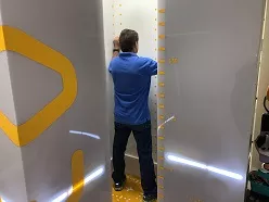
A young boy in an X-ray machine
Our 3D X-ray digital system provides low-dose, full-body, stereo-radiographic images of patients in a functional position. For instance, the system, described as a biplanar exam device, provides two perpendicular fan beams of X-rays and proprietary detectors. The proprietary detectors travel vertically while scanning the patient. In seconds, the EOS exam produces two simultaneous frontal and lateral, low-dose images or views of the whole body. It can also provide X-rays of particular body parts in the same short time. Similarly, your physician may select a particular position for the exam, including standing, bending, squatting, or sitting in a chair for a scoliosis X-ray.
Our 3D X-ray digital system represents the latest advancements in imaging technology. It provides a comprehensive and patient-centered approach to diagnosing and managing musculoskeletal conditions like Scoliosis. With its low-dose imaging, detailed 3D reconstructions, and advanced analysis capabilities, our system empowers us to provide personalized and effective care for patients with scoliosis and other spinal disorders.
What is the 3D X-ray Digital System?
Our 3D EOS X-ray digital machine contains the newest technology in scoliosis X-ray imaging. As such, it emits ‘low dose’ radiation that takes only 15 seconds to capture a full-standing image. Also, this machine achieves 80-90% less radiation exposure than older technology, according to the manufacturer. This significant reduction in exposure, often up to 10 times less than conventional X-ray systems, is a pivotal advancement in patient safety and diagnostic precision.
The EOS imaging system is renowned for its ability to deliver high-quality images with considerably lower radiation doses. This is especially critical for patients requiring frequent imaging, such as those with scoliosis. The Children’s Hospital of Wisconsin was an early adopter of the EOS’s efficacy and safety. It notes the substantial benefits over traditional methods.
With the growing number of U.S. facilities adopting EOS technology, it is becoming a more accessible option. Especially for those concerned with radiation exposure from conventional X-rays. Typically found in leading hospitals, EOS offers a safer diagnostic alternative required for accurate assessments.
This integration of cutting-edge technology and enhanced safety measures makes our 3D EOS X-ray machine an exemplary choice. It aids patients and healthcare providers aiming to reduce potential radiation risks while providing detailed and reliable imaging results.
Whole-body imaging:
Unlike conventional X-ray machines that focus on specific areas of the body, the 3D EOS system captures comprehensive, full-body images in both 2D and 3D. This allows healthcare providers to assess the entire spine and skeletal system in a single scan, providing a more complete understanding of the patient’s condition.
3D reconstruction and analysis:
The 3D EOS X-ray machine utilizes advanced software algorithms to reconstruct detailed three-dimensional models of the spine and surrounding structures. This allows for precise measurement and assessment of spinal curvature, alignment, and other parameters. The three-dimensional visualization provides enhanced diagnostic capabilities and aids in treatment planning.
Improved accuracy and precision:
The high-resolution images produced by the 3D EOS X-ray machine enable healthcare professionals to make more accurate measurements and assessments. This leads to improved surgical planning, better monitoring of treatment progress, and more precise evaluation of the outcomes.
Time-efficient and patient-friendly:
The 3D EOS X-ray machine offers rapid image acquisition, reducing the time required for patients to stay in the imaging room. Additionally, the open design of the machine provides a more comfortable and less restrictive experience for patients, particularly those with mobility challenges or claustrophobia.
Conclusion
Overall, the 3D EOS X-ray digital machine stands out for its advanced imaging capabilities, reduced radiation dose, comprehensive assessment of the spine and skeletal system, and enhanced precision in diagnosis and treatment planning. It represents a significant advancement in imaging technology for evaluating and managing various spinal conditions, including scoliosis. For adults with scoliosis, it is generally recommended to have an X-ray every five years. However, if symptoms begin to worsen, a more frequent X-ray is recommended to effectively monitor the condition.
Why is it Important to Get Low-Dose Radiation?
When discussing the impact of X-ray exposure on the human body, two primary theoretical models provide contrasting perspectives: the Linear threshold model and the Hormetic model.
Linear Threshold Model
The Linear threshold model, which is now considered somewhat outdated, traditionally posited a direct, no-threshold relationship between radiation exposure and the potential harm to human health. This model suggested that any amount of radiation, no matter how small, could lead to negative health effects without a safe exposure level.
Hormetic Model
On the other hand, the more recent Hormetic model introduces a new viewpoint. It suggests that lower doses of radiation might trigger beneficial biological responses. According to this model, such doses stimulate the body’s repair mechanisms, effectively preventing the cumulative damage usually associated with radiation exposure. This model challenges older views by proposing that small amounts of radiation could cause less harm than previously thought, and might even provide advantages.
These two models fundamentally differ in their assumptions about how radiation affects the body and the potential threshold levels considered safe for human exposure. The transition from the Linear threshold model to the Hormetic model marks a significant shift in understanding the biological impacts of X-ray exposure.
Results of Low Radiation Study
The study on radiation exposure in adolescent idiopathic scoliosis patients highlights a crucial concern: operative patients experience significantly higher radiation levels compared to those managed with braces or observation alone. Specifically, these patients are exposed to 8 to 14 times more radiation. This disparity underscores the importance of conservative treatments.
An alarming 78% of the annual radiation exposure for operative patients occurs during surgery, mainly due to intraoperative fluoroscopy and CT scans. This finding emphasizes the potential health risks associated with high radiation doses. Ultimately, the study reinforces the benefits of non-surgical interventions for scoliosis. By focusing on conservative treatments, patients can effectively manage their condition while minimizing harmful radiation exposure.
Recognizing the Symptoms of Scoliosis
Scoliosis is a condition characterized by an abnormal curve in the spine. Identifying its symptoms early on is crucial for effective treatment. Here are common signs to watch for if you suspect someone might have scoliosis:
Physical Imbalances
- Head Misalignment: The head does not center directly above the pelvis.
- Body Lean: Noticeable leaning of the body to one side.
- Ribcage and Hip Asymmetry: One side of the ribcage or hips may appear more prominent.
- Shoulder Disparities: Uneven shoulders, where one shoulder appears higher than the other, and shoulder blades that protrude.
Postural Changes
- Visible Curve in the Spine: In more severe cases, a curvature in the spine appears.
- Abnormal Walking Pattern: An uneven gait due to the body’s imbalanced alignment.
Pain and Discomfort
- Back Pain: Frequent or chronic pain in the back, which can vary in intensity.
- Muscle Tension: Tightness or spasms in the muscles surrounding the spine.
Understanding the different types of scoliosis is also vital for grasping the condition’s full scope:
Main Types of Scoliosis
- Idiopathic Scoliosis: The most prevalent form, making up about 80% of cases. It’s marked by an unexplained lateral spine curvature and often emerges during adolescence. Monitoring and a customized treatment plan are crucial for managing its development.
- Congenital Scoliosis: This form is present from birth due to spine development anomalies during fetal growth. These issues can lead to various spinal deformities and might require surgical intervention to avert further complications.
- Neuromuscular Scoliosis: Originating from neurological or muscular disorders like muscular dystrophy or cerebral palsy, this type can cause severe spinal deformities, impacting quality of life. Treatment typically involves bracing, physical therapy, and sometimes surgery to enhance mobility.
- Degenerative Scoliosis: Found in adults, this type arises from age-related spinal changes, such as osteoporosis or disc degeneration. It often results in back pain and stiffness, with treatment focusing on pain management and physical therapy, and occasionally surgery to improve spine alignment.
By recognizing both the symptoms and the types of scoliosis, individuals can seek timely medical advice and potentially halt the progression of this complex condition.
Diagnostic Steps for Young Patients
When a young patient presents with these symptoms, the path to diagnosis often begins with a routine physical exam. During this exam, healthcare professionals look for signs of abnormal spinal curvature. If scoliosis is suspected, it’s crucial to conduct a thorough physical examination.
- Medical History and Gait Observation: Doctors start by reviewing the patient’s medical history and observing their walking pattern, which can reveal a lot about the condition’s presence.
- Forward-Bend Test: Asymmetries are more noticeable when the patient bends forward, allowing for a clearer view of potential spinal curvatures.
- X-Ray Assessment: To confirm the diagnosis, a scoliosis X-ray is typically performed. This test helps in accurately measuring the degree of spinal curvature while carefully considering the need to minimize radiation exposure.
When to Seek Medical Advice
If you observe any of these symptoms, it’s crucial to consult a healthcare professional for a thorough examination. Early detection can significantly improve the management and treatment of scoliosis, ensuring a better quality of life.
Understanding these diagnostic processes and signs allows for timely intervention and effective management of scoliosis, particularly in adolescents, where the condition most commonly arises between the ages of 10 and 18.
What To Do If You Suspect Scoliosis
If you think you or a loved one might have scoliosis, it’s crucial to take immediate steps to address the condition. Here’s what you should do:
- Schedule a Doctor’s Appointment: First and foremost, book an appointment with a healthcare provider. A primary care physician can conduct an initial evaluation and refer you to a specialist if needed.
- Consult a Specialist: An orthopedic specialist or a spine specialist is well-equipped to diagnose and treat scoliosis. They can offer advanced diagnostics such as X-rays or MRI scans to confirm the presence and severity of scoliosis.
- Monitor Symptoms: Keep a close eye on any symptoms. Common signs include uneven shoulders, a visibly curved spine, and back pain. Documenting these can help your doctor make a more accurate diagnosis.
- Follow Recommended Treatments: Once diagnosed, adhere strictly to your treatment plan. This could range from physical therapy and braces to, in severe cases, surgical interventions.
- Seek Second Opinions: Don’t hesitate to get a second opinion to explore all possible treatment options. Consulting multiple specialists can provide a broader perspective on managing the condition.
- Stay Informed: Educate yourself about scoliosis. Websites like this one, the Scoliosis Research Society, and the National Scoliosis Foundation offer valuable resources.
By following these steps, people can take a proactive approach to managing scoliosis effectively. Always prioritize professional medical advice to ensure the best outcomes.
Understanding Idiopathic Scoliosis in Adults Versus Children
Idiopathic scoliosis is a condition characterized by abnormal curvature of the spine, and it affects both children and adults. However, the manifestation and progression differ significantly between these age groups.
Key Differences
1. Origin:
- Children: Idiopathic scoliosis in children often emerges during growth spurts in adolescence. The cause is unknown, which is why it’s termed “idiopathic.”
- Adults: In adults, idiopathic scoliosis typically represents a continuation of scoliosis that was either undiagnosed, untreated, or ineffectively treated during childhood. Over time, the curve can worsen due to factors like aging, osteoporosis, or degenerative changes.
2. Symptoms:
- Children: Symptoms in children may not be immediately obvious. Often, signs like uneven shoulders or hips prompt further examination.
- Adults: Adults may experience more pronounced symptoms, including back pain, stiffness, posture changes, and visible spinal curvature. Pain is more common due to the added stress on the spine and surrounding muscles over the years of compensation.
3. Progression:
- Children: The curvature often progresses rapidly during periods of growth. If detected early, bracing and other interventions can effectively halt or reduce further curvature.
- Adults: Progression in adults tends to appear gradual but can accelerate due to degenerative spinal disorders. Management focuses on alleviating pain and preventing further deterioration.
4. Treatment Approaches:
- Children: Treatments for children typically include observation, bracing, and, in severe cases, surgery. Early intervention can significantly alter the course of the condition.
- Adults: Adults are often treated with a combination of physical therapy, pain management, medications, and occasionally surgery. The goals are to manage symptoms and improve quality of life rather than curative measures.
Summary
While idiopathic scoliosis in both adults and children involves spinal curvature of unknown origin, adults often face more complex and symptomatic conditions due to the condition persisting and evolving over time. Recognizing these differences is crucial for effective management of scoliosis at various life stages.
Every case of scoliosis is different, which is why a customized treatment approach is so important. Treatment plans must address the specifics of each patient and their condition. This is where X-ray results become invaluable, as they provide detailed insights into the unique curvature and progression of the spine.
A tailored approach ensures that the nuances of each individual’s scoliosis are considered, enhancing the potential for successful outcomes. This personalized strategy is vital not only for addressing current symptoms but also for preventing further progression, particularly in adult patients who may experience more pronounced symptoms over time.
How Does the Location of a Scoliosis Curvature Affect Its Progression?
The progression of scoliosis is closely linked to where the curvature is found along the spine. Certain areas are more prone to noticeable changes over time, influencing both the rate of progression and treatment approach.
Key Factors
- Thoracic Curves: Curvatures in the thoracic region, which includes the upper and mid-back, often progress more rapidly to thoracic scoliosis. This is due to the unique anatomical and biomechanical demands placed on this part of the spine.
- Lumbar Curves: Curves located in the lower back might progress to lumbar scoliosis differently. These tend to develop at a slower pace but can significantly impact posture and balance.
- Double Curves: Sometimes, scoliosis presents as a double curve, such as an S-shape. The interaction between the two curves can affect overall progression, often necessitating more complex management strategies.
Understanding the precise location of a spinal curvature is crucial. It helps healthcare professionals predict how scoliosis might develop over time and tailor treatments effectively to each individual’s needs.
How Many People in the US Are Affected by Scoliosis?
Scoliosis, a condition characterized by an abnormal curvature of the spine, impacts a substantial segment of the US population. Estimates suggest that between 6 million and 9 million Americans live with scoliosis. This condition is typically identified during childhood, although its severity varies widely among individuals.
While some individuals with mild scoliosis may not experience noticeable symptoms, others might suffer from back pain, spinal weakness, and even difficulties with breathing or maintaining a sitting posture for extended periods. Additionally, it’s worth noting that females are significantly more likely than males—by a factor of eight—to develop severe curvature that necessitates medical intervention.
Why is Scoliosis X-ray Imaging Conducted?
 An X-ray will display your bones and how they relate to each other. In addition, an X-ray helps your doctor determine if a patient has a fracture of the spine, an infection, or a tumor. For over 100 years, doctors have continued to use X-rays to check bone alignments and to see whether certain shadows appear out of alignment. With this tool, doctors can get clues about the health of the spine. If your doctor thinks your problem emanates from degeneration of the spine, X-rays can determine if the space between your vertebrae has decreased, if bone spurs exist, or if hypertrophy (enlargement) of the facet joints exists.
X-rays remain the gold standard for diagnosing, assessing, and monitoring scoliosis due to their precision and reliability. They provide crucial measurements, such as the Cobb angle, to evaluate how a scoliotic spine deviates from normal alignment. This measurement is essential for understanding the degree of curvature and tailoring an effective treatment plan. Moreover, X-rays can reveal spinal rotation and help determine if the condition meets the criteria for structural scoliosis.
While there are other methods like MRIs that doctors use, X-rays are unmatched in their accuracy, accessibility, and cost-effectiveness when it comes to scoliosis. MRIs can show no evidence of significant abnormalities other than scoliosis itself, offering a comprehensive view to ensure no other underlying conditions are present.
Correctly interpreting X-rays
To specifically diagnose idiopathic scoliosis, doctors look for predictable patterns such as right thoracic (middle back) scoliosis or left thoracolumbar (mid and low-back) scoliosis. “If no other related condition is present to suggest other scoliosis causes—such as neuromuscular or congenital—the doctor will diagnose idiopathic scoliosis,” says Dr. Hostin. This exclusion is crucial because it helps to pinpoint idiopathic scoliosis accurately, distinguishing it from other types of scoliosis that may require different treatment approaches.
It’s important to note that the practitioner interpreting the X-ray should have scoliosis-specific training and experience. This expertise ensures the 3-dimensional complexities of the condition are fully understood, allowing for the most effective treatment planning.
Diagnosis Verification
By combining X-ray and MRI results, doctors can assess both the alignment of the bones and any changes in the soft tissues. This thorough evaluation process ensures that idiopathic scoliosis is diagnosed accurately, allowing for a tailored treatment plan.
Specifically, in the context of scoliosis, X-rays serve an indispensable role. They enable physicians to measure Cobb angles, identify spinal and rib deformations, and closely monitor changes in curvature. This is crucial during adolescence, a phase when scoliosis can rapidly progress during growth spurts. Despite concerns about radiation exposure, it’s important to note that, according to the Health Physics Society, there is no conclusive evidence of radiation causing harm at the levels patients receive from diagnostic X-ray exams. Furthermore, a study specific to scoliosis radiology states, “Patients undergoing scoliosis radiography receive effective doses that are low in comparison with other types of radiographic examination.”
Understanding these details underscores the necessity of X-rays not just for general spinal health but crucially in managing and monitoring scoliosis.
Characteristics Required for a Structural Scoliosis Diagnosis
When diagnosing structural scoliosis, there are certain key criteria that doctors look for. Here’s a breakdown of what healthcare professionals typically look for:
- Abnormal Sideways Curvature: The spine must exhibit a sideways curve that deviates from the normal alignment. This curvature isn’t just slight; it needs to have a serious curve to qualify as scoliosis.
- Minimum Curve Size: To receive a structural scoliosis diagnosis, the curvature must surpass a specific degree. Generally, this means a curve greater than 10 degrees as measured on an X-ray.
- Spinal Rotation: Unlike non-structural forms, structural scoliosis includes a rotational component. This means that the vertebrae not only bend sideways but also rotate, often causing rib prominence or a more noticeable physical appearance.
- Permanence of the Curve: The curvature and rotation are not flexible. When postural adjustments or external force are applied, the curve and rotation remain unchanged, indicating a more permanent structural issue.
Recognizing these signs can assist in distinguishing structural scoliosis from less severe spinal issues. Accurate diagnosis requires thorough clinical evaluation, often involving imaging techniques like X-rays.
Conducting a Scoliosis X-ray
Having an X-ray taken is much like having your photograph taken. For instance, it is a quick and painless procedure. You will lie very still on a table or stand very still and hold certain positions while pictures are taken of your spine. Sometimes X-ray technicians will ask that you stand or sit in different positions. For example, the technician can take the X-ray while you bend forward (flexion), and another while you straighten your spine (extension). For example, doctors refer to this as a “flexion-extension” view of the spine. Moreover, these X-rays are compared to see if there is extra movement between the vertebrae, a condition called segmental instability.
For Patients in Pain
Our X-ray system will take the X-ray really fast, within 15 seconds. Also, the system can take 3D weight-bearing images that provide your physician with views of your spine or limb anatomy that are not available with 2D X-rays, but become critical for diagnosing and treating complex orthopedic and spine conditions.
What are the Limitations of Scoliosis X-ray Imaging?
X-rays do not show the soft tissues-nerves, discs, and ligaments. Today, many tests can show the soft tissues much more clearly, so doctors do not always have to rely on X-rays. However, X-rays provide a good starting point in evaluating the spine.
Understanding Scoliosis with 2D X-Ray Images
Scoliosis is a complex, three-dimensional condition, manifesting not just with a sideward curve but also with a rotational twist of the spine. To truly comprehend this intricate disorder, specialists rely on two-dimensional X-rays.
The Role of 2D X-Rays in Scoliosis Assessment
While traditional X-rays offer only flat, 2D representations, experts use multiple views to piece together a full, 3D understanding of the spine. This involves taking X-ray images from various perspectives—side-to-side, front-to-back, and reverse. These multiple images help construct a comprehensive overview, revealing vital nuances such as rotation and tilt.
Why Expertise Matters
Effective scoliosis treatment hinges on accurately deciphering these X-ray images. Specialists are skilled in extracting critical measurements that go beyond the basic Cobb angle, such as assessing the spine’s twist and tilt. This ensures a precise diagnosis, enabling tailored treatment strategies.
Ensuring Accurate Diagnosis
Without the expertise to interpret multiple 2D images as a cohesive 3D model, doctors can miss vital details, potentially undermining treatment efficacy. Hence, it’s crucial to consult healthcare providers specializing in scoliosis who can effectively translate these images into actionable insights.
In summary, although X-rays are inherently two-dimensional, with the right expertise, they become powerful tools in understanding and managing the three-dimensional intricacies of scoliosis.
What X-ray Techniques Can Further Diagnose Patient Conditions?
Beyond the commonly used lateral bending scoliosis X-rays, healthcare professionals can further assess a patient’s spinal condition using additional X-ray techniques. One valuable method includes sagittal X-rays, which provide a side view of the spine. These images are particularly useful for examining the sagittal profile of the spine, offering detailed insight into its alignment and curvature.
To evaluate the flexibility of the spine, the technician will take sagittal images while the patient bends to the left and then to the right. This approach helps determine the range of motion and flexibility within the spine, crucial for devising appropriate treatment plans. Such comprehensive imaging techniques ensure a more accurate assessment of the spinal condition, aiding in more targeted and effective interventions.
Comprehensive Monitoring Techniques
- Regular X-ray Evaluation: Essential for tracking scoliosis progression, X-rays help measure the Cobb angle and identify spinal and rib deformations. By comparing images taken at different intervals, healthcare providers can assess how the condition evolves and adjust treatment plans accordingly.
- Physical Examination: A thorough physical exam is vital for evaluating the patient’s overall health, detecting changes in spinal curvature, and assessing treatment effectiveness. During these exams, healthcare providers look for signs of asymmetry and posture changes, which are indicators of scoliosis progression.
- Scoliometer Measurements: This non-invasive tool measures the degree of spinal curvature. Used alongside X-rays, scoliometer measurements provide a comprehensive assessment of scoliosis, helping track changes in curvature and detect potential progression.
Two types of X-ray views are commonly necessary for monitoring scoliosis. First, a full spine view is captured, which can display either an anterior-to-posterior (AP) or a posterior-to-anterior (PA) view. This provides a comprehensive look at the spine from the front or back. The second required view is the sagittal view, which offers a detailed side perspective of the spine. These two X-ray angles together help healthcare professionals accurately assess the curvature and progression of scoliosis.
By integrating these varied methods, healthcare providers ensure a thorough and nuanced approach to monitoring and managing scoliosis, leading to more effective and personalized treatment strategies.
Getting Up-to-date X-rays for Adolescents
Up-to-date X-rays are crucial for effective scoliosis treatment in both adolescents and adults, though the reasons vary between the two groups. For adolescents, whose bodies are still growing, timely X-rays allow physicians to monitor the progression of spinal curvature accurately. Without current X-rays, there’s a risk of basing treatment on outdated information, which can lead to ineffective management or even worsening of the condition. Identifying specific curve patterns through recent X-rays is essential for making informed decisions, particularly when utilizing specialized treatment methods like the Schroth Method and Chêneau bracing.
Adolescents diagnosed with scoliosis often require frequent X-ray monitoring, especially during growth spurts. These periods of rapid growth can lead to quick changes or progression in the curvature of the spine. It’s crucial to obtain timely X-rays to ensure that any changes are accurately tracked and addressed promptly. While it’s important to minimize unnecessary radiation exposure, relying on outdated X-rays can result in inadequate treatment, which is not advisable for a growing adolescent.
Comprehensive Monitoring Approaches
Monitoring scoliosis progression involves more than just X-rays. Regular check-ups with healthcare providers are essential to track changes in spinal curvature and detect potential complications. The frequency of monitoring is influenced by the severity of scoliosis, the patient’s age, and the chosen treatment approach. Here’s a closer look at the methods involved:
- Physical Examinations: These are vital for assessing overall health and detecting changes in spinal curvature. Healthcare providers look for signs of asymmetry and changes in posture, which are critical indicators of scoliosis progression.
- Scoliometer Measurements: This non-invasive tool measures the degree of spinal curvature and is often used alongside X-rays. Regular scoliometer assessments help track changes and provide a comprehensive picture of the condition.
By incorporating these varied methods, healthcare providers can ensure a holistic approach to scoliosis management, tailored to the individual needs of the patient. This comprehensive monitoring helps in making informed decisions to adjust treatment plans effectively and mitigate the risk of progression.
Adult X-ray Frequency
In grown-ups, how often X-rays are done can stretch to longer gaps since their spines do not change as quickly. Still, when starting a new plan of care, new X-rays matter. An image that is three or four years old will not help a doctor, especially if the patient has pain. New scans show the spine’s state more clearly, which is key for making a care plan that meets the needs of now, not the past.
What are the Risks?
Our X-ray tool gives less radiation dose to our patients. For instance, the EOS X-ray system gives 50% to 85% fewer rays than old digital X-ray tools and 95% less dose than CT scans. For those who need many X-rays to track their spine, the EOS system is the best choice. It gives fast, safe, and sharp images.
Most patients who get X-rays will never get enough rays to fear cancer. Only those who get many X-rays over the years may face that risk. Also, doctors guard kids and young adults from rays to the sex glands when treating spine bends or other spine problems. This is due to rays that may harm sperm or eggs. During X-rays, it is easy to guard these parts with a lead apron or lead cloth.
Now, with new health tools, going to places like the Southwest Scoliosis and Spine Institute that use new digital X-ray tools can help patients. These new tools aim to cut ray dose, which makes them the best choice for those who need many scans. Always ask what tools your scan site uses and pick one that gives the most safety with the least radiation.
To keep the tech safe, it’s best to stand six feet or more from the X-ray unit when you can. This cuts the rays the tech gets.
How Does the Scoliosis 3DC® Program Aim to Reduce X-ray Exposure While Managing Scoliosis?
At Scoliosis 3DC®, cutting X-ray dose is a key part of our full plan for care. We put patient safety first while making sure tests are right, and care stays on track.
Baseline and Follow-Up X-Rays
To begin, we acquire precise baseline Cobb angle measurements via a scoliosis series x-ray of the full spine. This includes:
- An anterior-to-posterior (AP) or posterior-to-anterior (PA) view of the entire spine.
- A sagittal (side) view of the spine.
For subsequent check-ups, typically only a single AP or PA view is needed, reducing the overall number of X-rays required during treatment.
Tailored Frequency of X-Rays
The frequency of follow-up X-rays is customized based on each patient’s specific needs, including the severity of the curve and bone maturity, as indicated by the Risser sign. Understanding scoliosis severity is crucial for tailoring these follow-ups. Here’s how scoliosis is categorized based on Cobb angle measurements:
- Mild scoliosis: Cobb angle measurement of between 10 and 25 degrees
- Moderate scoliosis: Cobb angle measurement of between 25 and 40 degrees
- Severe scoliosis: Cobb angle measurement of 40+ degrees
- Very-severe scoliosis: Cobb angle measurement of 80+ degrees
By categorizing scoliosis in this way, healthcare providers can better determine the appropriate monitoring and intervention strategies, ensuring each patient receives personalized care that aligns with their unique condition.
Safety Precautions
When X-rays are necessary, we implement several safety measures to further reduce radiation exposure:
- Patients stand at least six feet away from the X-ray unit.
- Lead aprons are used to shield sensitive areas such as the breasts and pelvic region from radiation.
Use of Advanced Technology
We advocate for the use of modern digital X-ray equipment, which emits significantly less radiation than traditional radiography methods. This advanced technology is generally available at our facilities, providing an additional layer of safety for our patients.
Patient Advocacy
Informed patients are empowered patients. We educate everyone in our program to advocate for themselves regarding radiation safety during X-rays. This includes understanding the necessity of shielding and the benefits of digital X-ray technology.
By incorporating these measures, Scoliosis 3DC® ensures high-quality care while prioritizing the reduction of X-ray exposure, thus promoting better long-term health for our patients.
__________________
The medical content on this page has been carefully reviewed and approved for accuracy by the Southwest Scoliosis and Spine Institute’s qualified healthcare professionals, including our board-certified physicians and Physician Assistants. Our team ensures that all information reflects the latest evidence-based practices and meets rigorous standards of medical accuracy, with oversight from our expert spine doctors to guarantee reliability for our patients.
If you or a loved one suffers from spinal pain, you owe it to yourself to call Southwest Scoliosis and Spine Institute at 214-556-0555 to make an appointment.