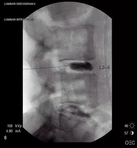
Discogram
A discogram refers to injecting a dye into your spine and then taking X-ray pictures of the discs in the spine. To see the discs, the doctor injects a dye into the center of the injured disc(s). Upon the doctor examining the pictures, the discs will appear visible on X-ray film and a fluoroscope (special X-ray TV screen.
Why use a Discogram?
This test will determine which disc has structural damage and where the pain originates from. If a disc has begun to rupture and if it has tears in the tough outer ring (the annulus), a discogram will identify these problems. Also, by injecting fluid into the disc to increase pressure, the doctor can tell if it generates pain.
Normal discs, and even those that are severely degenerated, do not usually cause pain. Before surgery, a discogram helps the doctor know the location of the problem and the type of operation needed.
The Benefits
- Identification of Pain Source: A discogram can help identify if a particular disc causes pain. By injecting a contrast dye into the disc, the procedure can reproduce the patient’s pain symptoms, and identify a particular disc as the pain generator. Therefore, this information can provide valuable data for doctors to make treatment decisions and determine the most appropriate course of action.
- Surgical Planning: Discograms can aid in surgical planning for individuals with severe disc-related problems. If conservative treatments do not provide sufficient relief, a discogram can help identify which specific discs cause pain. Consequently, this information is useful for surgeons in deciding whether a particular disc should be repaired, removed, or replaced during surgical intervention.
- Accurate Diagnosis: For individuals with complex or atypical pain patterns, a discogram can help provide a more accurate diagnosis. Furthermore, it can distinguish between multiple discs as potential sources of pain, and rule out discs not generating symptoms. This can help avoid unnecessary treatments or interventions and focus on addressing the root cause of the pain.

How to Conduct a Discogram
Prior to the operation, medication will help you relax. Then the doctor will apply a local anesthetic to numb the area of the back. Upon numbing the area, the doctor injects the dye into the nucleus pulposus (the very center of the intervertebral dick). Most importantly, the doctor uses a fluoroscope to see your spine, and the needle gets inserted into the correct disc space. Then X-rays and a CT scan take pictures to see a cross-section of the disc. The procedure lasts about 40 minutes
In addition, once the needle gets placed inside the damaged disc, the doctor places a small amount of fluid to increase pressure on the disc. If this test causes pain similar to your back or leg pain, it reveals that the disc causes the pain.
Limitations of a Discogram?
Unfortunately, the discogram does not show the bones or nerves very well-only inside the intervertebral disc. Doctors do not frequently use this test; however, if an MRI fails to find a problem, disc doctors will recommend it. Doctors also rely on the discogram when considering disc surgery.
The risks?
The risks associated with a discogram include infection inside the disc and an allergic reaction to the dye. Discograms require X-rays, which use radiation. In large doses, radiation can increase the risk of cancer. The vast majority of patients who undergo X-rays will never get enough radiation to worry about cancer. Only doctors whose patients must get hundreds of X-rays over many years become concerned. This test has more risks associated with it than most. This provides the reason that doctors prefer to use “noninvasive” tests first, such as MRI and CT scans.
If you or a loved one suffers from spinal pain, you owe it to yourself to call Southwest Scoliosis and Spine Institute at 214-556-0555 to make an appointment.


