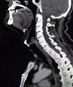
MYLEOGRAM OF THE SPINE

A myelogram of the spine, a diagnostic test, helps visualize the spinal canal. It also might help identify conditions affecting the spinal cord, discs, and nerves in the spine. This test combines an X-ray with an injected dye that helps to illuminate the spine.

Take the Scoliosis Quiz
Take the Pain Quiz

Cervical Spine CT Scan

Myelogram of the Spine

A myelogram, an older test, examines the spinal canal and spinal cord. The doctor injects a special dye into the spinal sac that will indicate any abnormalities on X-rays. Before there were CT and MRI scans, the myelogram was the best test to determine the cause of pressure on the spinal cord or spinal nerves. Today the myelogram only gets used for very special purposes, such as for complicated revision spine surgeries. If you have a herniated disc, it rarely becomes the first test used.

Why is it done?

The dye used during a myelogram outlines the spinal cord and nerve roots. This helps your doctor see any unusual indentations or abnormal shapes in the spinal cord. Anything that pushes into the nerves shows up as an indentation into the spinal sac. This indentation could originate from a herniated or bulging disc, a tumor, or an injury to the spinal nerve roots. For patients who have metal plates and screws in their spine, the myelogram becomes the first choice because metal prevents MRI scans.  

Before a Myelography

Before undergoing myelography, it’s crucial to inform your physician about specific medical conditions and situations to ensure your safety and the procedure’s efficacy. Make sure to discuss the following:

Existing Medical Conditions

  • Allergies: Highlight any allergies you have, particularly to contrast media, latex, or any other substances.
  • Heart Issues: Mention if you have angina or any other heart problems.
  • Epilepsy: If you suffer from epilepsy, this is important to disclose.

Medications and Treatments

  • Blood Thinners: Let your doctor know if you are taking any anticoagulant medications.
  • Diabetes: Inform the physician about your diabetes and any medications you are taking for it.

Current Physical Conditions

  • Kidney Disease: Report any kidney issues, as these can affect the use of contrast media.
  • Difficulty Urinating: Mention if you have any problems with urination, which can be relevant during the procedure.

Special Circumstances

  • Pregnancy: Notify if you are pregnant or suspect you might be.
  • Breastfeeding: If you are breastfeeding, this is essential information for your physician.
  • Mobility Issues: Discuss any difficulties you have lying on your stomach or remaining still for extended periods.

By providing this information, you help your healthcare provider take the necessary precautions and tailor the procedure to your individual needs, ensuring the best possible outcome.

How Should You Prepare for a Myelography Test?

Preparing for a myelography test requires careful planning and adherence to specific guidelines to ensure your safety and the accuracy of the test results. Follow these preparation steps:

  • Arrange Transportation: Ensure you have someone to drive you both to the facility and back home post-test. The test can affect your ability to drive safely.

Pre-Appointment Checklist

Previous Medical Records:

  • Bring Relevant Scans: Gather copies of past imaging studies such as X-rays, CT scans, or MRIs. These may be requested for the neuroradiologist to compare with the new images.

Fasting Guidelines:

  • No Solid Food: Do not consume any solid food after midnight before your appointment.
  • Clear Liquids: Drinking clear liquids (like water) is usually permitted, but always check with your physician for any specific restrictions.

Medication Instructions

  • Continue Medications Carefully: On the day of the test, you may need to take your prescribed medications with clear liquids, unless otherwise instructed by your doctor. Confirm these details in advance to avoid any complications.

Personal Belongings

  • Avoid Valuables: It’s advised to leave jewelry and other valuables at home to prevent any loss or interference during the procedure.

General Tips

  • Follow Doctor’s Instructions: Always adhere strictly to the guidelines provided by your healthcare provider. Each patient may receive personalized instructions based on their individual medical condition.

By following these steps, you can help ensure a smooth and successful myelography test experience.

Who Typically Performs a Myelography Test?

A myelography test is generally conducted by a specialized medical professional known as a neuroradiologist. This type of radiologist has advanced training and expertise in imaging techniques specifically focused on the nervous system and spine.

How to conduct a Myelogram of the spine

The doctor must perform a spinal tap to inject dye into the spinal sac. The dye mixes with the spinal fluid so that it will show up on X-rays. You will lie on a tilting table while multiple X-rays are taken to show the flow of the dye through the spine. The myelogram is usually combined with a CT scan to get a better view of the spine in cross-section and to check the health of the bones and nerves.

Step-by-Step Procedure

  • Preparation:
    After changing into a gown, you will be positioned on your stomach or side for the injection.
  • The injection area of the skin is cleansed thoroughly.

Numbing the Area:

  • A numbing medication is injected to help make the procedure more comfortable.
  • You might feel a sting or gentle pressure as the needle is inserted. Some patients report sharp but momentary pain.

Injection of Contrast Medium:

  • Once the area is numb, the contrast medium (dye) is injected into the spinal sac.
  • The dye mixes with the spinal fluid to enhance visibility on X-rays.

Imaging:

  • The neuroradiologist uses fluoroscopy—real-time video X-ray—to ensure the contrast medium flows into the targeted area of the spine.
  • Multiple X-rays are taken to show the flow of the dye through the spine.
  • The myelogram is usually combined with a CT scan to get a better view of the spine in cross-section and to assess the health of the bones and nerves.

By following these detailed steps, the myelogram provides a comprehensive view of the spine, aiding in the diagnosis and treatment of spinal conditions.

What are the limitations?

A myelogram does not show the soft tissues. It shows only the bones and the spinal fluid where the dye has mixed with the fluid.

What are the risks?

Because the myelogram requires a spinal tap, there are more risks associated with it than most other tests. This provides one reason that doctors prefer to use “noninvasive” tests first, such as MRI and CT scans. Although the risks associated with a spinal tap are rare, they include meningitis (infection of the spinal fluid), spinal headache, and allergic reaction to the dye. However, the needle might cause bleeding around the spinal sac. Meanwhile, the myelogram requires X-rays, which use radiation, and large doses of radiation can increase the risk of cancer. Finally, the vast majority of patients who have X-rays taken will never get enough radiation to worry about cancer. Only patients who have large numbers of X-rays — hundreds over many years — need to be concerned.

Common Side Effects

In addition to the risks mentioned above, there are also common side effects that patients might experience. These include:

  • Headache
  • Aches or discomfort in the arms or legs
  • Nausea
  • Vomiting
  • Dizziness

Most patients do not experience any side effects, and when they do occur, they usually disappear within 24 hours.

What Aftercare is Required Following a Myelography Procedure?

After a myelography procedure, precise aftercare steps are essential to ensure proper recovery. Here’s what you need to know:
Immediate Recovery:

  • You’ll be taken to a recovery area where you’ll need to rest for up to four hours. This period is crucial for monitoring your initial response to the procedure.

Positioning During Recovery:

  • Some patients may be positioned with their heads elevated. This can help mitigate certain side effects and ensure comfort.

Monitoring:

  • The radiology staff will closely observe you throughout this recovery period to ensure that no immediate complications arise.

Discharge Instructions:

  • Activity Restrictions: Avoid strenuous activities for the first 24 hours. Also, refrain from bending movements for 48 hours to prevent complications.
  • Hydration: It’s important to drink plenty of fluids, such as water or juice. This helps flush the contrast dye from your system.
  • Breastfeeding Considerations: If you are breastfeeding, you might be advised to give your baby formula for a short duration after the test to ensure safety.

By adhering to these aftercare instructions, you can help ensure a smoother recovery after your myelography.

When to Contact a Physician

If side effects persist longer than 24 hours, or if you develop a fever over 100.4 degrees F, it is important to contact your physician, the hospital, or the radiology clinic where the test was performed. This ensures that any potential complications are addressed promptly.

Balancing Risks and Benefits

While the risks and side effects associated with myelography can be concerning, it’s important to balance these against the benefits of the test. Myelography can provide critical information that other tests may not be able to, which can be vital for accurate diagnosis and treatment planning. Always discuss any concerns with your healthcare provider to make an informed decision about the procedure.

If you or a loved one suffers from spinal pain, you owe it to yourself to call Southwest Scoliosis and Spine Institute at 214-556-0555 to make an appointment.

Cookie Preferences