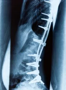
The surgeons at the Southwest Scoliosis and Spine Institute use screws and rods to keep vertebra in place while healing
 Hardware for Spine Fusion
Southwest Scoliosis and Spine Institute’s board-certified, fellowship-trained orthopedic physicians, Richard Hostin, MD, Devesh Ramnath, MD, Ishaq Syed, MD, Shyam Kishan, MD, and Kathryn Wiesman, MD, have years of experience treating thousands of patients with complex spine conditions. In addition, they possess extensive knowledge and experience with “rods and pedicle screws” that are needed for each surgery. They spend lots of time studying and participating in research to ensure their patients get the correct procedure for their condition.
Pedicle Screws for Spine Fusion Rationale
The right combination of metal screws and rods (hardware) creates a solid “brace” that holds the vertebrae in place. These devices stop the movement from occurring between the vertebrae. These metal devices give more stability to the fusion site and allow the patient to get out of bed much sooner.
Spine Fusion Procedure
Spinal fusion permanently connects two or more vertebrae in your spine, eliminating motion between them. Spinal fusion involves techniques to increase healing. The use of pedicle screws improves spinal fusion rates from approximately 60% to 90%. Many surgeons also believe that pedicle screws enhance patient recovery because they provide immediate stability for the spine and early mobilization for the patient.
Initially, the safety and effectiveness of using pedicle screws were brought to the FDA for approval. Therefore, the FDA studied the use of these screws and approved them for use in the lower (lumbar) spine for specific conditions.
Also, the technique for placing the pedicle screws in the patient requires a steep learning curve. And only surgeons like Dr. Hostin, an experienced surgeon utilizing pedicle screws for decades, should use them. Importantly, a pedicle screw provides a means of gripping a spinal segment. Further, the screws themselves act as firm anchor points that can then connect with a stabilizing rod.
Implementation
Surgeons place Pedicle screws with the help of pedicle bone on the back of the spinal column. The screw inserts through the pedicle and into the vertebral body, one on each side. The screws grab into the bone of the vertebral body, and thus, provide a solid hold on the vertebra. In addition, the breakage rate is reduced to about one in 1,000 with modern pedicle screws. Once the doctor places the screws, they attach these screws to metal rods that connect all the screws together. When the doctor bolts together and tightens everything, it will create a stiff metal frame that holds the vertebrae. This process will help in healing. The bone graft gets a place around the back of the vertebrae.
After the bone graft grows, the screws and rods are no longer needed for stability. Also, you can remove it safely through subsequent back surgery. However, most surgeons do not recommend removal unless the pedicle screws cause discomfort for the patient (5% to 10% of cases).
Subsequently, 350 physicians of the North American Spine Society conducted an analysis of 2,500 patients. These doctors found a very low complication rate using pedicle screws in spinal fusion surgery. Further, information discloses only about a one in 1,000 chance of nerve root damage, and a 2% to 3% chance of infection.
If you or a loved one suffers from spinal pain, you owe it to yourself to call Southwest Scoliosis and Spine Institute at 214-556-0555 to make an appointment.


