Lumbar Discectomy: A Comprehensive Guide
 Back pain, mainly in the lower back, is a common problem that can greatly harm daily life. When basic care does not ease pain from a slipped disc in the lower spine, a lumbar discectomy often serves as the main surgery. This surgery is one of the most commonly done back ops in the world, known for its strong track record in easing nerve pain and bringing back movement. This page looks at what a lumbar discectomy is, how surgeons do it, and why they use it. It also gives clear points on how to get ready, heal, avoid risks, and know the likely results.
Lumbar Discectomy
A lumbar discectomy is a back surgery to take out part or all of a slipped or torn disc in the lower spine, the part with five bones (L1 to L5). The disc, made of a soft core (nucleus) wrapped by a tough outer ring (annulus), works like a pad between the bones. When the soft core breaks through a tear in the ring, the slipped disc can press on the nearby nerves or the spinal cord, which may cause pain, numbness, or weak strength. A lumbar discectomy takes out the slipped part to ease the squeeze on nerves and cut pain.
The word “discectomy” means “disc” (the spinal disc) plus “ectomy” (take out). In most cases, surgeons take out just the slipped part and leave the rest of the disc to keep the spine steady. Some types, like microdiscectomy or less-cut discectomy, use new tools to cut less tissue.
Lumbar Discectomy and how surgeons use it
Surgeons use a Lumbar discectomy primarily to treat conditions involving disc herniation in the lower back that do not respond to non-surgical interventions like physical therapy, medications, or injections. Its key indications include:
Lumbar Disc Herniation:
- When the nucleus pulposus bulges or ruptures through the annulus, it presses on the spinal nerve roots (most commonly at L4-L5 or L5-S1). This often causes sciatica—radiating pain, tingling, or numbness down one or both legs.
Radiculopathy:
- Nerve root irritation or compression leading to symptoms like leg weakness, burning sensations, or loss of reflexes (e.g., diminished knee or ankle reflex).
Cauda Equina Syndrome (Rare Emergency):
- A severe herniation compressing the cauda equina (nerve bundle at the spinal cord’s end), causing bowel or bladder dysfunction, saddle anesthesia (numbness around the groin), and leg weakness. This requires urgent surgery.
Chronic Pain or Disability:
- When herniation-related symptoms persist for 6-12 weeks despite conservative care, they disrupt daily activities or work.
Surgeons do not use a Lumbar discectomy for degenerative disc disease without herniation, spinal stenosis without nerve compression, or non-specific lower back pain without neurological symptoms. Its focus is on relieving nerve pressure rather than addressing structural spinal issues like arthritis or misalignment.
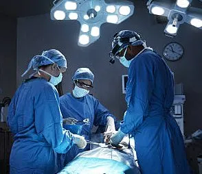
How do Surgeons Perform Lumbar Discectomy?
Surgeons perform Lumbar discectomy using different techniques—open discectomy, microdiscectomy, or minimally invasive approaches, each tailored to the patient’s condition and the surgeon’s expertise. Below is a detailed breakdown of today’s most common method, microdiscectomy, which replaces traditional open surgery due to its precision and reduced recovery time.
Preoperative Preparation
- Evaluation: Patients undergo a physical exam, MRI or CT scan to confirm the herniation’s location and severity, and sometimes an electromyography (EMG) to assess nerve damage.
- Medical Clearance: Blood tests, heart monitoring (e.g., EKG), and medication reviews ensure surgical safety.
- Instructions: Patients fast for 8-12 hours pre-surgery, stop certain medications (e.g., blood thinners), and arrange post-operative support.
Surgical Procedure
Anesthesia:
- Doctors put Patients to sleep with general anesthesia, rendering them unconscious and pain-free throughout the procedure.
Positioning:
- The patient lies face-down (prone) on a specialized table with padding to maintain spinal alignment and allow X-ray guidance (fluoroscopy).
Incision:
- Surgeons make a small incision (1-2 cm for microdiscectomy, 3-5 cm for open surgery) over the affected disc level (e.g., L4-L5), guided by preoperative imaging.
Exposure:
- Muscle Retraction: Surgeons move back muscles (paraspinals) gently aside using retractors, not cutting, to access the spine. In microdiscectomy, a tubular retractor minimizes tissue disruption.
- Laminotomy: A small portion of the lamina (bony arch of the vertebra) is removed to expose the disc and nerve root. This step is less extensive than in a laminectomy.
Disc Removal:
- Nerve Protection: Surgeons lift or retract the compressed nerve root using a nerve hook or retractor.
- Herniated Material Extraction: Using microsurgical tools (e.g., forceps, rongeurs) under a microscope or magnifying loupes, the surgeon removes the protruding nucleus pulposus. The goal is to decompress the nerve without destabilizing the spine.
- Annulus Inspection: The surgeon will clear any loose disc fragments, but the intact disc remains in place to maintain cushioning.
Closure:
- The surgeons then close the incision with sutures or staples, and the incision is covered by a sterile dressing. Surgeons do not need to use hardware (e.g., screws, rods), unlike in spinal fusion.
Duration:
- The procedure lasts 1-2 hours, depending on complexity (e.g., single vs. multi-level herniation).
Variations
- Open Discectomy: A Larger incision and more muscle dissection are used in complex cases or when microscopes aren’t available.
- Endoscopic Discectomy: Uses a tiny camera (endoscope) through an even smaller incision, further reducing trauma.
- Laser Discectomy: Rarely used, employs a laser to vaporize herniated tissue, though evidence of efficacy is limited.
Postoperative Process
- Immediate Recovery: Patients wake in a recovery room, monitored for 1-2 hours. Most microdiscectomy patients go home the same day (outpatient), while open surgery may require a 1-2 night hospital stay.
- Pain Management: Mild incision pain is managed with oral painkillers (e.g., acetaminophen, NSAIDs); nerve pain often subsides quickly.
- Activity: Walking is encouraged within hours, but bending, twisting, or lifting (>5-10 lbs) is avoided for 2-6 weeks.
- Follow-Up: Doctors remove stitches (if non-dissolvable) in 7-14 days; physical therapy may start at 2-4 weeks to strengthen the back.
Risks and Complications of Lumbar Discectomy
Though generally safe (success rate >90%), lumbar discectomy carries potential risks:
- Infection: 1-2% risk at the incision site or deeper (discitis).
- Nerve Damage: Rare (<1%), but can cause persistent weakness or numbness.
- Recurrent Herniation: 5-10% chance of the same disc re-herniating, especially if activity resumes too soon.
- Dural Tear: Leakage of spinal fluid (1-3%), repairable during surgery, but may delay recovery.
- Bleeding: Minimal with microdiscectomy, higher with open surgery.
Outcomes and Effectiveness
- Pain Relief: 85-95% of patients experience a significant reduction in leg pain (sciatica) within days to weeks, per studies in the Journal of Neurosurgery.
- Function: Most resume normal activities within 4-6 weeks, with full recovery by 3 months.
- Back Pain: Localized lower back pain may persist if degenerative changes (e.g., arthritis) are present, though nerve symptoms improve.
- Long-Term: Success depends on avoiding re-injury and maintaining spinal health through exercise and posture.
When is it Not Appropriate?
Lumbar discectomy does not work for:
- Non-herniated disc issues (e.g., degenerative disc disease without nerve compression).
- Spinal instability (e.g., spondylolisthesis) may require fusion.
- Patients with minimal symptoms or those improving with conservative care.
Conclusion
Lumbar discectomy is a focused, strong surgery to ease nerve compression from a slipped disc in the lower back. By taking out the disc part that presses on nerves with fine steps like a microdiscectomy, it treats leg pain, weak strength, and numbness, often with fast healing and little break in life. Used most for long-term or bad nerve pain, it shows back surgery’s mix of skill and safe care. For those with nonstop low back disc pain, this surgery gives a sure way to get back to moving and ease, if paired with good care after surgery and healthy life steps. Parents should seek care for a child with a spine curve at the top care sites.
At the Southwest Scoliosis and Spine Institute, we focus on Scoliosis Diagnosis, Treatment, & Care for our Patients. Our fellowship-trained, board-certified expert orthopedic scoliosis surgeons, Richard Hostin, MD, Devesh Ramnath, MD, Ishaq Syed, MD, Shyam Kishan, MD, and Kathryn Wiesman, MD, specialize in all types of spine conditions, deformities, and scoliosis pain. Our group has skills in the diagnosis and care of all back problems. The Southwest Scoliosis and Spine Institute, with offices in Dallas, Plano, and Frisco, Texas, is here for all spine patients.
This spine group brings in a team of back doctors and pain care experts. The Institute also gives us access to top imaging tools and care paths. For one, it uses the newest proven steps to help patients heal fast. Each patient also gets a care plan made to fit their own needs and health history. In the end, the choice of where to get care will rest on many things, but for those who want the best, parents should look first to the Southwest Scoliosis and Spine Institute.
____________________
Citation: Science Direct – Surgical Discectomy for Lumbar Disc Herniation
The medical content on this page has been carefully reviewed and approved for accuracy by the Southwest Scoliosis and Spine Institute’s qualified healthcare professionals, including our board-certified physicians and Physician Assistants. Our team ensures that all information reflects the latest evidence-based practices and meets rigorous standards of medical accuracy, with oversight from our expert spine doctors to guarantee reliability for our patients.
If you or your loved one is suffering from back pain from a spinal condition, there is hope. We can help. Call Southwest Scoliosis and Spine Institute at 214-556-0555 to make an appointment today.
Recent Comments